| کد مقاله | کد نشریه | سال انتشار | مقاله انگلیسی | نسخه تمام متن |
|---|---|---|---|---|
| 6291081 | 1302476 | 2013 | 12 صفحه PDF | دانلود رایگان |
عنوان انگلیسی مقاله ISI
Increased action of triclabendazole (TCBZ) in vitro against a TCBZ-resistant isolate of Fasciola hepatica following its co-incubation with the P-glycoprotein inhibitor, R(+)-verapamil
دانلود مقاله + سفارش ترجمه
دانلود مقاله ISI انگلیسی
رایگان برای ایرانیان
کلمات کلیدی
موضوعات مرتبط
علوم زیستی و بیوفناوری
ایمنی شناسی و میکروب شناسی
انگل شناسی
پیش نمایش صفحه اول مقاله

چکیده انگلیسی
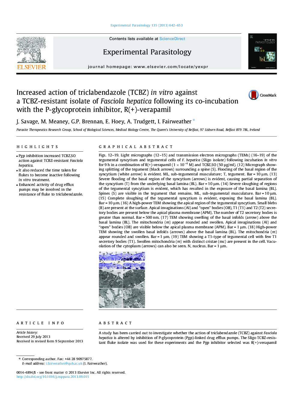
Figs. 12-19. Light micrographs (12-15) and transmission electron micrographs (TEMs) (16-19) of the tegumental syncytium and tegumental cells of F. hepatica (Sligo isolate) following incubation in vitro for 9 h in a combination of R(+)-verapamil (1 Ã 10â4 M) and TCBZ.SO (50 μg/ml). (12) Micrograph showing splitting of the tegument (black arrows) surrounding a spine (S). Flooding of the basal region of the syncytium (white arrow) is evident. ML, sub-tegumental musculature; T, tegument. Bar = 10 μm. (13) Severe flooding of the basal region of the syncytium (arrows) is evident, causing partial separation of the syncytium (T) from the underlying basal lamina (BL). Bar = 10 μm. (14) Severe sloughing of regions of the tegumental syncytium is evident, which has resulted in the exposure of the basal lamina (BL). Spines (S) are visible in the tegument that remains. ML, sub-tegumental musculature. Bar = 10 μm. (15) Complete sloughing of the tegumental syncytium is evident, exposing the basal lamina (BL). Bar = 10 μm. (16) A high-power TEM showing the apical region of the tegumental syncytium. Small blebs (B) are present at the surface. Apical invaginations (AI) and “open” bodies (OB), T1 (T1) and T2 (T2) secretory bodies are present below the apical plasma membrane (APM). The number of T2 secretory bodies is greater than normal. Bar = 500 nm. (17) TEM showing swelling of the basal infolds (arrow) above the basal lamina (BL). The mitochondria (m) appear rounded and swollen. Apical invaginations (AI) and “open” bodies (OB) are visible below the apical plasma membrane (APM). Bar = 1 μm. (18) High-power TEM showing the swollen basal infolds (arrows) above the basal lamina (BL). The mitochondria (m) appear rounded and swollen. Bar = 1 μm. (19) TEM showing a T1-type of tegumental cell with few T1 secretory bodies (T1). Swollen mitochondria (m) with distinct cristae (mc) are present in the cell. Vacuolation of the cytoplasm (arrows) can also be seen. N, nucleus. Bar = 1 μm.
ناشر
Database: Elsevier - ScienceDirect (ساینس دایرکت)
Journal: Experimental Parasitology - Volume 135, Issue 3, November 2013, Pages 642-653
Journal: Experimental Parasitology - Volume 135, Issue 3, November 2013, Pages 642-653
نویسندگان
J. Savage, M. Meaney, G.P. Brennan, E. Hoey, A. Trudgett, I. Fairweather,