| کد مقاله | کد نشریه | سال انتشار | مقاله انگلیسی | نسخه تمام متن |
|---|---|---|---|---|
| 9902209 | 1545794 | 2005 | 12 صفحه PDF | دانلود رایگان |
عنوان انگلیسی مقاله ISI
Flow cytometric determination of mitochondrial membrane potential changes during apoptosis of T lymphocytic and pancreatic beta cell lines: Comparison of tetramethylrhodamineethylester (TMRE), chloromethyl-X-rosamine (H2-CMX-Ros) and MitoTracker Red 580 (
دانلود مقاله + سفارش ترجمه
دانلود مقاله ISI انگلیسی
رایگان برای ایرانیان
کلمات کلیدی
موضوعات مرتبط
علوم زیستی و بیوفناوری
بیوشیمی، ژنتیک و زیست شناسی مولکولی
بیوتکنولوژی یا زیستفناوری
پیش نمایش صفحه اول مقاله

چکیده انگلیسی
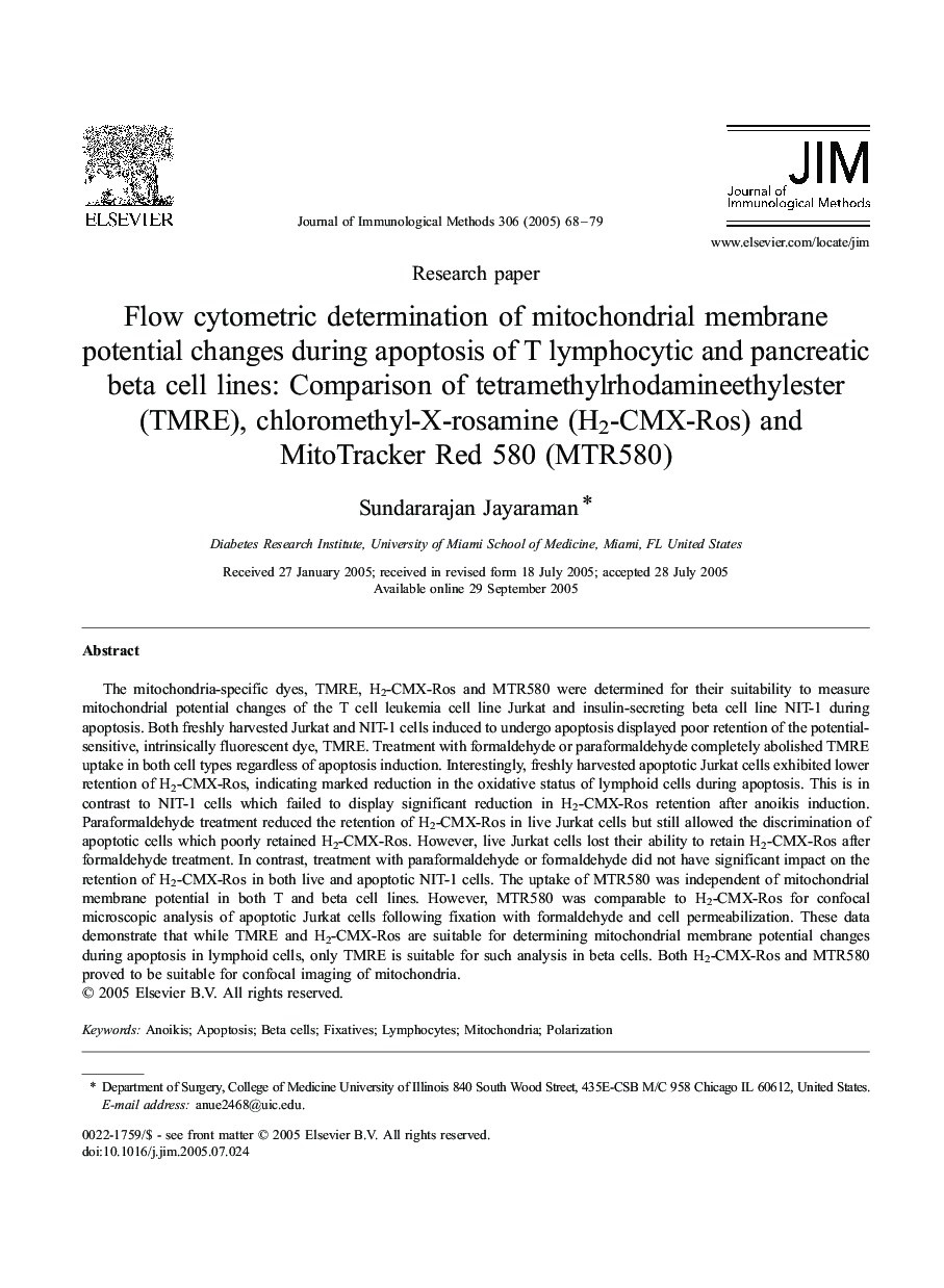
The mitochondria-specific dyes, TMRE, H2-CMX-Ros and MTR580 were determined for their suitability to measure mitochondrial potential changes of the T cell leukemia cell line Jurkat and insulin-secreting beta cell line NIT-1 during apoptosis. Both freshly harvested Jurkat and NIT-1 cells induced to undergo apoptosis displayed poor retention of the potential-sensitive, intrinsically fluorescent dye, TMRE. Treatment with formaldehyde or paraformaldehyde completely abolished TMRE uptake in both cell types regardless of apoptosis induction. Interestingly, freshly harvested apoptotic Jurkat cells exhibited lower retention of H2-CMX-Ros, indicating marked reduction in the oxidative status of lymphoid cells during apoptosis. This is in contrast to NIT-1 cells which failed to display significant reduction in H2-CMX-Ros retention after anoikis induction. Paraformaldehyde treatment reduced the retention of H2-CMX-Ros in live Jurkat cells but still allowed the discrimination of apoptotic cells which poorly retained H2-CMX-Ros. However, live Jurkat cells lost their ability to retain H2-CMX-Ros after formaldehyde treatment. In contrast, treatment with paraformaldehyde or formaldehyde did not have significant impact on the retention of H2-CMX-Ros in both live and apoptotic NIT-1 cells. The uptake of MTR580 was independent of mitochondrial membrane potential in both T and beta cell lines. However, MTR580 was comparable to H2-CMX-Ros for confocal microscopic analysis of apoptotic Jurkat cells following fixation with formaldehyde and cell permeabilization. These data demonstrate that while TMRE and H2-CMX-Ros are suitable for determining mitochondrial membrane potential changes during apoptosis in lymphoid cells, only TMRE is suitable for such analysis in beta cells. Both H2-CMX-Ros and MTR580 proved to be suitable for confocal imaging of mitochondria.
ناشر
Database: Elsevier - ScienceDirect (ساینس دایرکت)
Journal: Journal of Immunological Methods - Volume 306, Issues 1â2, 30 November 2005, Pages 68-79
Journal: Journal of Immunological Methods - Volume 306, Issues 1â2, 30 November 2005, Pages 68-79
نویسندگان
Sundararajan Jayaraman,