| کد مقاله | کد نشریه | سال انتشار | مقاله انگلیسی | نسخه تمام متن |
|---|---|---|---|---|
| 10097649 | 1609881 | 2005 | 6 صفحه PDF | دانلود رایگان |
عنوان انگلیسی مقاله ISI
Assessment of talus deformity by three-dimensional MRI in congenital clubfoot
دانلود مقاله + سفارش ترجمه
دانلود مقاله ISI انگلیسی
رایگان برای ایرانیان
کلمات کلیدی
موضوعات مرتبط
علوم پزشکی و سلامت
پزشکی و دندانپزشکی
رادیولوژی و تصویربرداری
پیش نمایش صفحه اول مقاله

چکیده انگلیسی
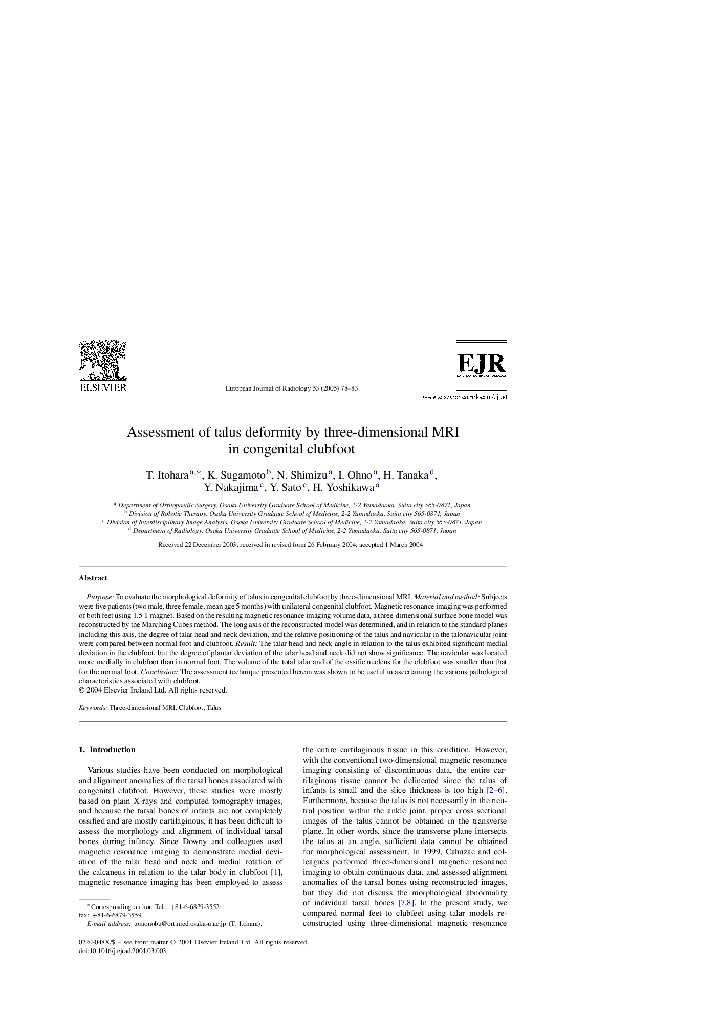
Purpose: To evaluate the morphological deformity of talus in congenital clubfoot by three-dimensional MRI. Material and method: Subjects were five patients (two male, three female, mean age 5 months) with unilateral congenital clubfoot. Magnetic resonance imaging was performed of both feet using 1.5Â T magnet. Based on the resulting magnetic resonance imaging volume data, a three-dimensional surface bone model was reconstructed by the Marching Cubes method. The long axis of the reconstructed model was determined, and in relation to the standard planes including this axis, the degree of talar head and neck deviation, and the relative positioning of the talus and navicular in the talonavicular joint were compared between normal foot and clubfoot. Result: The talar head and neck angle in relation to the talus exhibited significant medial deviation in the clubfoot, but the degree of plantar deviation of the talar head and neck did not show significance. The navicular was located more medially in clubfoot than in normal foot. The volume of the total talar and of the ossific nucleus for the clubfoot was smaller than that for the normal foot. Conclusion: The assessment technique presented herein was shown to be useful in ascertaining the various pathological characteristics associated with clubfoot.
ناشر
Database: Elsevier - ScienceDirect (ساینس دایرکت)
Journal: European Journal of Radiology - Volume 53, Issue 1, January 2005, Pages 78-83
Journal: European Journal of Radiology - Volume 53, Issue 1, January 2005, Pages 78-83
نویسندگان
T. Itohara, K. Sugamoto, N. Shimizu, I. Ohno, H. Tanaka, Y. Nakajima, Y. Sato, H. Yoshikawa,