| کد مقاله | کد نشریه | سال انتشار | مقاله انگلیسی | نسخه تمام متن |
|---|---|---|---|---|
| 10097654 | 1609881 | 2005 | 10 صفحه PDF | دانلود رایگان |
عنوان انگلیسی مقاله ISI
MR imaging features of foot involvement in ankylosing spondylitis
دانلود مقاله + سفارش ترجمه
دانلود مقاله ISI انگلیسی
رایگان برای ایرانیان
کلمات کلیدی
موضوعات مرتبط
علوم پزشکی و سلامت
پزشکی و دندانپزشکی
رادیولوژی و تصویربرداری
پیش نمایش صفحه اول مقاله

چکیده انگلیسی
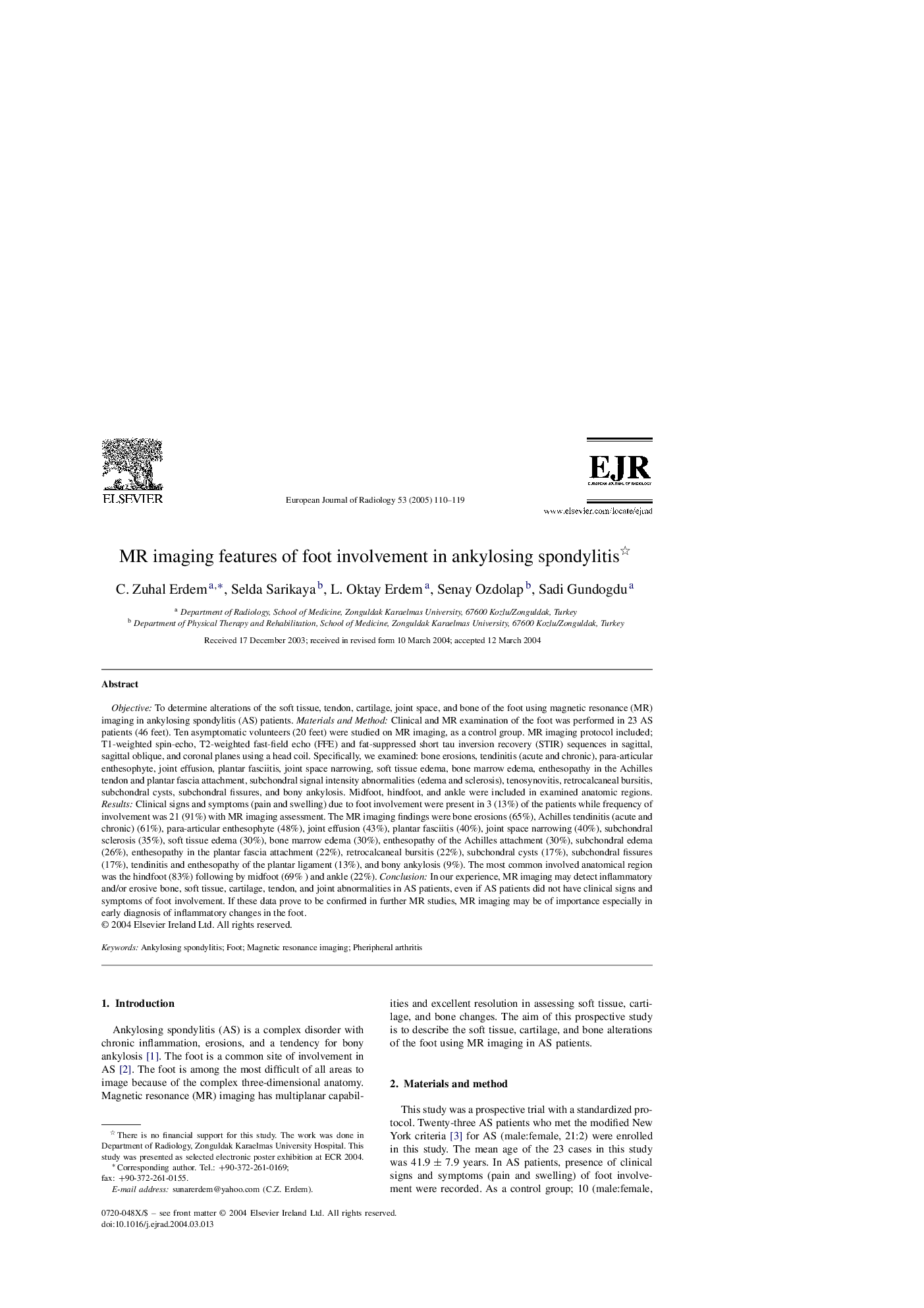
Objective: To determine alterations of the soft tissue, tendon, cartilage, joint space, and bone of the foot using magnetic resonance (MR) imaging in ankylosing spondylitis (AS) patients. Materials and Method: Clinical and MR examination of the foot was performed in 23 AS patients (46 feet). Ten asymptomatic volunteers (20 feet) were studied on MR imaging, as a control group. MR imaging protocol included; T1-weighted spin-echo, T2-weighted fast-field echo (FFE) and fat-suppressed short tau inversion recovery (STIR) sequences in sagittal, sagittal oblique, and coronal planes using a head coil. Specifically, we examined: bone erosions, tendinitis (acute and chronic), para-articular enthesophyte, joint effusion, plantar fasciitis, joint space narrowing, soft tissue edema, bone marrow edema, enthesopathy in the Achilles tendon and plantar fascia attachment, subchondral signal intensity abnormalities (edema and sclerosis), tenosynovitis, retrocalcaneal bursitis, subchondral cysts, subchondral fissures, and bony ankylosis. Midfoot, hindfoot, and ankle were included in examined anatomic regions. Results: Clinical signs and symptoms (pain and swelling) due to foot involvement were present in 3 (13%) of the patients while frequency of involvement was 21 (91%) with MR imaging assessment. The MR imaging findings were bone erosions (65%), Achilles tendinitis (acute and chronic) (61%), para-articular enthesophyte (48%), joint effusion (43%), plantar fasciitis (40%), joint space narrowing (40%), subchondral sclerosis (35%), soft tissue edema (30%), bone marrow edema (30%), enthesopathy of the Achilles attachment (30%), subchondral edema (26%), enthesopathy in the plantar fascia attachment (22%), retrocalcaneal bursitis (22%), subchondral cysts (17%), subchondral fissures (17%), tendinitis and enthesopathy of the plantar ligament (13%), and bony ankylosis (9%). The most common involved anatomical region was the hindfoot (83%) following by midfoot (69% ) and ankle (22%). Conclusion: In our experience, MR imaging may detect inflammatory and/or erosive bone, soft tissue, cartilage, tendon, and joint abnormalities in AS patients, even if AS patients did not have clinical signs and symptoms of foot involvement. If these data prove to be confirmed in further MR studies, MR imaging may be of importance especially in early diagnosis of inflammatory changes in the foot.
ناشر
Database: Elsevier - ScienceDirect (ساینس دایرکت)
Journal: European Journal of Radiology - Volume 53, Issue 1, January 2005, Pages 110-119
Journal: European Journal of Radiology - Volume 53, Issue 1, January 2005, Pages 110-119
نویسندگان
C.Zuhal Erdem, Selda Sarikaya, L.Oktay Erdem, Senay Ozdolap, Sadi Gundogdu,