| کد مقاله | کد نشریه | سال انتشار | مقاله انگلیسی | نسخه تمام متن |
|---|---|---|---|---|
| 10691044 | 1019580 | 2016 | 8 صفحه PDF | دانلود رایگان |
عنوان انگلیسی مقاله ISI
Color Doppler Ultrasound in Diagnosis and Assessment of Carotid Body Tumors: Comparison with Computed Tomography Angiography
ترجمه فارسی عنوان
سونوگرافی داپلر رنگی در تشخیص و ارزیابی تومورهای بدن کاروتید: مقایسه با آنژیوگرافی توموگرافی کامپیوتری
دانلود مقاله + سفارش ترجمه
دانلود مقاله ISI انگلیسی
رایگان برای ایرانیان
کلمات کلیدی
تومور بدن کاروتید، تشخیص، سونوگرافی رنگ داپلر، آنژیوگرافی کامپیوتری توموگرافی،
موضوعات مرتبط
مهندسی و علوم پایه
فیزیک و نجوم
آکوستیک و فرا صوت
چکیده انگلیسی
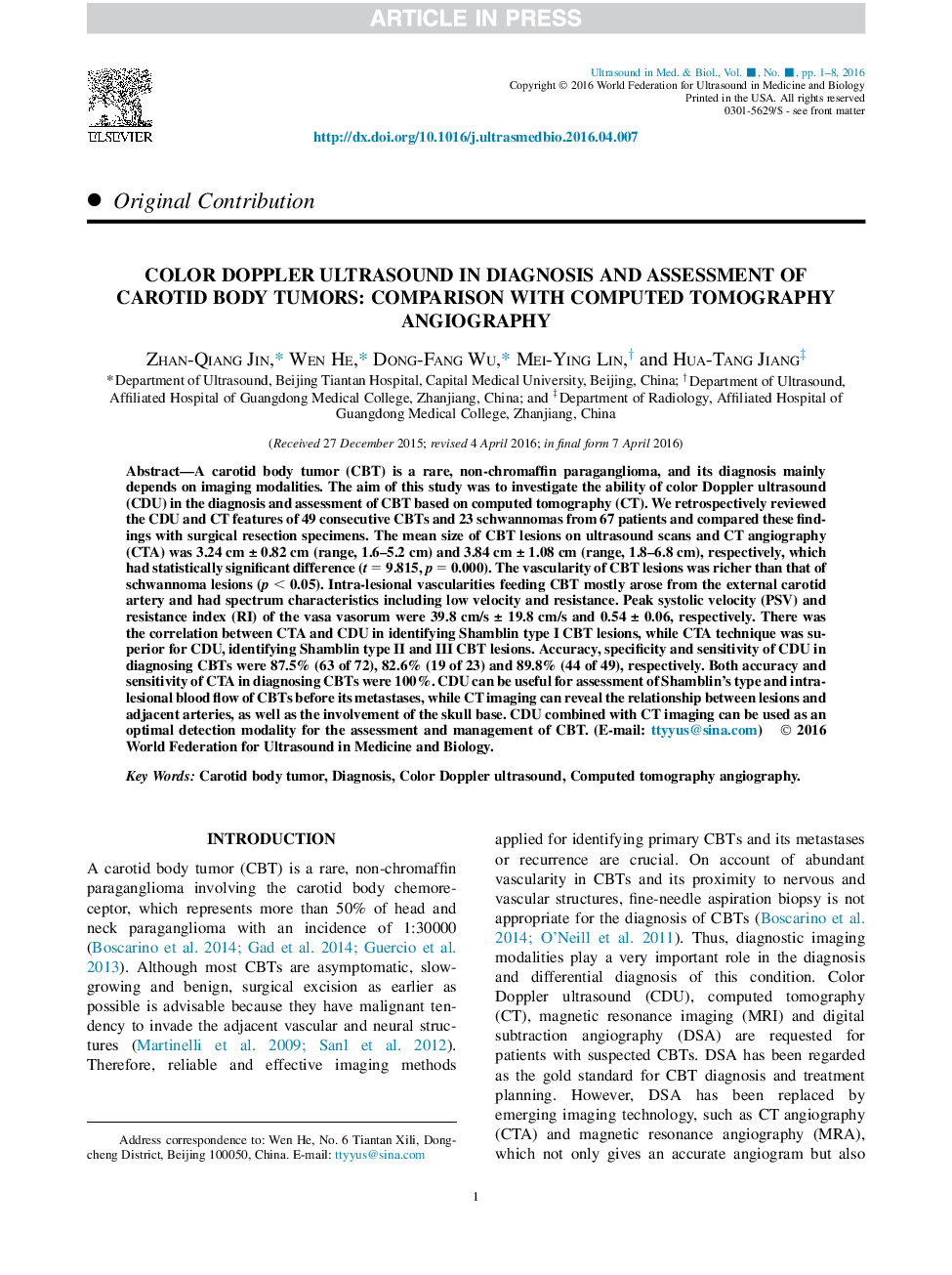
A carotid body tumor (CBT) is a rare, non-chromaffin paraganglioma, and its diagnosis mainly depends on imaging modalities. The aim of this study was to investigate the ability of color Doppler ultrasound (CDU) in the diagnosis and assessment of CBT based on computed tomography (CT). We retrospectively reviewed the CDU and CT features of 49 consecutive CBTs and 23 schwannomas from 67 patients and compared these findings with surgical resection specimens. The mean size of CBT lesions on ultrasound scans and CT angiography (CTA) was 3.24 cm ± 0.82 cm (range, 1.6-5.2 cm) and 3.84 cm ± 1.08 cm (range, 1.8-6.8 cm), respectively, which had statistically significant difference (t = 9.815, p = 0.000). The vascularity of CBT lesions was richer than that of schwannoma lesions (p < 0.05). Intra-lesional vascularities feeding CBT mostly arose from the external carotid artery and had spectrum characteristics including low velocity and resistance. Peak systolic velocity (PSV) and resistance index (RI) of the vasa vasorum were 39.8 cm/s ± 19.8 cm/s and 0.54 ± 0.06, respectively. There was the correlation between CTA and CDU in identifying Shamblin type I CBT lesions, while CTA technique was superior for CDU, identifying Shamblin type II and III CBT lesions. Accuracy, specificity and sensitivity of CDU in diagnosing CBTs were 87.5% (63 of 72), 82.6% (19 of 23) and 89.8% (44 of 49), respectively. Both accuracy and sensitivity of CTA in diagnosing CBTs were 100%. CDU can be useful for assessment of Shamblin's type and intra-lesional blood flow of CBTs before its metastases, while CT imaging can reveal the relationship between lesions and adjacent arteries, as well as the involvement of the skull base. CDU combined with CT imaging can be used as an optimal detection modality for the assessment and management of CBT.
ناشر
Database: Elsevier - ScienceDirect (ساینس دایرکت)
Journal: Ultrasound in Medicine & Biology - Volume 42, Issue 9, September 2016, Pages 2106-2113
Journal: Ultrasound in Medicine & Biology - Volume 42, Issue 9, September 2016, Pages 2106-2113
نویسندگان
Zhan-Qiang Jin, Wen He, Dong-Fang Wu, Mei-Ying Lin, Hua-Tang Jiang,
