| کد مقاله | کد نشریه | سال انتشار | مقاله انگلیسی | نسخه تمام متن |
|---|---|---|---|---|
| 10691828 | 1019611 | 2014 | 7 صفحه PDF | دانلود رایگان |
عنوان انگلیسی مقاله ISI
Quantification of Carotid Plaque Neovascularization Using Contrast-Enhanced Ultrasound With Histopathologic Validation
ترجمه فارسی عنوان
کوانتومی نئواساکولاریزاسیون پلاک کاروتید با استفاده از سونوگرافی پیشرفته کنتراست با اعتبار سنجی هیستوپاتولوژیک
دانلود مقاله + سفارش ترجمه
دانلود مقاله ISI انگلیسی
رایگان برای ایرانیان
کلمات کلیدی
موضوعات مرتبط
مهندسی و علوم پایه
فیزیک و نجوم
آکوستیک و فرا صوت
چکیده انگلیسی
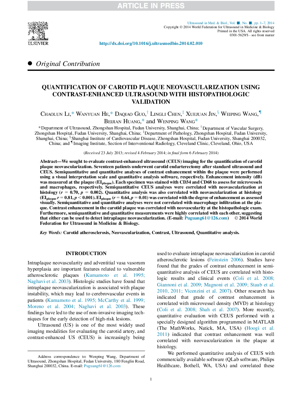
We sought to evaluate contrast-enhanced ultrasound (CEUS) imaging for the quantification of carotid plaque neovascularization. Seventeen patients underwent carotid endarterectomy after standard ultrasound and CEUS. Semiquantitative and quantitative analyses of contrast enhancement within the plaque were performed using a visual interpretation scale and quantitative analysis software, respectively. Enhancement intensity (dB) was measured at the plaque (EIplaque). Each specimen was stained with CD34 and CD68 to assess for microvessels and macrophages, respectively. Semiquantitative CEUS analyses were correlated with neovascularization at histology (r = 0.70, p = 0.002). Quantitative analysis was also correlated with neovascularization at histology (EIplaquer = 0.81, p < 0.001). EIplaque (r = 0.64, p = 0.01) was correlated with the degree of enhancement as assessed visually. Semiquantitative and quantitative analyses were not correlated with macrophage infiltration at the plaque. Contrast enhancement in the carotid plaque was correlated with neovascularity at the histopathologic exam. Furthermore, semiquantitative and quantitative measurements were highly correlated with each other, suggesting that either can be used to detect intraplaque neovascularization.
ناشر
Database: Elsevier - ScienceDirect (ساینس دایرکت)
Journal: Ultrasound in Medicine & Biology - Volume 40, Issue 8, August 2014, Pages 1827-1833
Journal: Ultrasound in Medicine & Biology - Volume 40, Issue 8, August 2014, Pages 1827-1833
نویسندگان
Chaolun Li, Wanyuan He, Daqiao Guo, Lingli Chen, Xuejuan Jin, Weiping Wang, Beijian Huang, Wenping Wang,
