| کد مقاله | کد نشریه | سال انتشار | مقاله انگلیسی | نسخه تمام متن |
|---|---|---|---|---|
| 1760991 | 1019629 | 2009 | 12 صفحه PDF | دانلود رایگان |
عنوان انگلیسی مقاله ISI
Mean Volume Flow Estimation in Pulsatile Flow Conditions
دانلود مقاله + سفارش ترجمه
دانلود مقاله ISI انگلیسی
رایگان برای ایرانیان
کلمات کلیدی
موضوعات مرتبط
مهندسی و علوم پایه
فیزیک و نجوم
آکوستیک و فرا صوت
پیش نمایش صفحه اول مقاله

چکیده انگلیسی
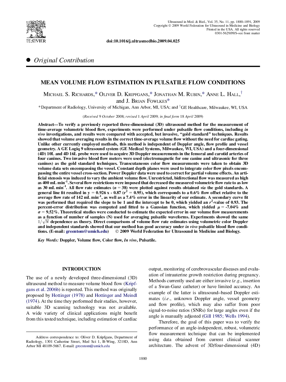
To verify a previously reported three-dimensional (3D) ultrasound method for the measurement of time-average volumetric blood flow, experiments were performed under pulsatile flow conditions, including in vivo investigations, and results were compared with accepted, but invasive, “gold standard” techniques. Results showed that volume averaging results in the correct time-average volume flow without the need for cardiac gating. Unlike other currently employed methods, this method is independent of Doppler angle, flow profile and vessel geometry. A GE Logiq 9 ultrasound system (GE Medical Systems, Milwaukee, WI, USA) and a four-dimensional (4D) 10L and 4D 16L probe were used to acquire 3D Doppler measurements in the femoral and carotid arteries of four canines. Two invasive blood flow meters were used (electromagnetic for one canine and ultrasonic for three canines) as the gold standard techniques. Transcutaneous color flow measurements were taken to obtain 3D volume data sets encompassing the vessel. Constant depth planes were used to integrate color flow pixels encompassing the entire vessel cross-section. Power Doppler data were used to correct for partial volume effects. An artificial stenosis was induced to vary the ambient volume flow. Unrestricted, bidirectional flow was measured as high as 400 mL min-1. Several flow restrictions were imposed that decreased the measured volumetric flow rate to as low as 30 mL min-1. All flow rate estimates (n = 38) were plotted against results obtained via the gold standards. A general line fit resulted in y = 0.926 x - 0.87 (r2 = 0.95), which corresponds to a 0.6% flow offset relative to the average flow rate of 142 mL min-1, as well as a 7.4% error in the linearity of our estimate. A secondary curve fit was performed that required the slope to be 1 and the intercept to be 0, which yielded an r2-value of 0.93. The percent-error distribution was computed and fitted to a Gaussian function, which yielded μ = -7.04% and Ï = 9.52%. Theoretical studies were conducted to estimate the expected error in our volume flow measurements as a function of number of samples (N) used for averaging pulsatile waveforms. Experiments showed the same 1/N dependence as theory. Direct comparisons of volume flow rate estimates using volumetric color Doppler and independent standards showed that our method has good accuracy under in vivo pulsatile blood flow conditions. (E-mail: [email protected])
ناشر
Database: Elsevier - ScienceDirect (ساینس دایرکت)
Journal: Ultrasound in Medicine & Biology - Volume 35, Issue 11, November 2009, Pages 1880-1891
Journal: Ultrasound in Medicine & Biology - Volume 35, Issue 11, November 2009, Pages 1880-1891
نویسندگان
Michael S. Richards, Oliver D. Kripfgans, Jonathan M. Rubin, Anne L. Hall, J. Brian Fowlkes,