| کد مقاله | کد نشریه | سال انتشار | مقاله انگلیسی | نسخه تمام متن |
|---|---|---|---|---|
| 2734660 | 1147672 | 2008 | 7 صفحه PDF | دانلود رایگان |
عنوان انگلیسی مقاله ISI
Imagerie des sarcomes utérins
دانلود مقاله + سفارش ترجمه
دانلود مقاله ISI انگلیسی
رایگان برای ایرانیان
کلمات کلیدی
موضوعات مرتبط
علوم پزشکی و سلامت
پزشکی و دندانپزشکی
انفورماتیک سلامت
پیش نمایش صفحه اول مقاله

چکیده انگلیسی
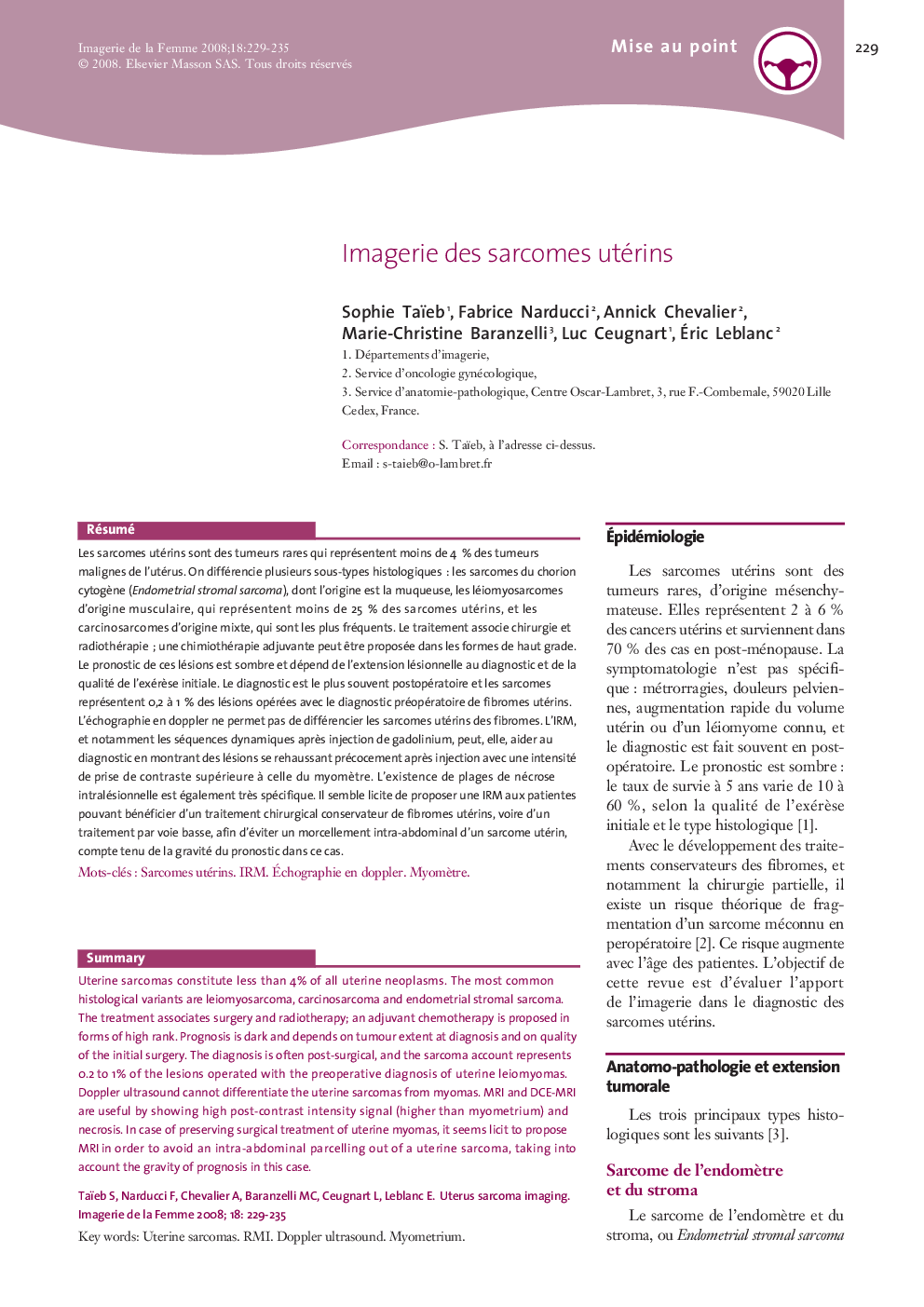
Uterine sarcomas constitute less than 4% of all uterine neoplasms. The most common histological variants are leiomyosarcoma, carcinosarcoma and endometrial stromal sarcoma. The treatment associates surgery and radiotherapy; an adjuvant chemotherapy is proposed in forms of high rank. Prognosis is dark and depends on tumour extent at diagnosis and on quality of the initial surgery. The diagnosis is often post-surgical, and the sarcoma account represents 0.2 to 1% of the lesions operated with the preoperative diagnosis of uterine leiomyomas. Doppler ultrasound cannot differentiate the uterine sarcomas from myomas. MRI and DCE-MRI are useful by showing high post-contrast intensity signal (higher than myometrium) and necrosis. In case of preserving surgical treatment of uterine myomas, it seems licit to propose MRI in order to avoid an intra-abdominal parcelling out of a uterine sarcoma, taking into account the gravity of prognosis in this case.
ناشر
Database: Elsevier - ScienceDirect (ساینس دایرکت)
Journal: Imagerie de la Femme - Volume 18, Issue 4, December 2008, Pages 229-235
Journal: Imagerie de la Femme - Volume 18, Issue 4, December 2008, Pages 229-235
نویسندگان
Sophie Taïeb, Fabrice Narducci, Annick Chevalier, Marie-Christine Baranzelli, Luc Ceugnart, Ãric Leblanc,