| کد مقاله | کد نشریه | سال انتشار | مقاله انگلیسی | نسخه تمام متن |
|---|---|---|---|---|
| 3171092 | 1199838 | 2011 | 10 صفحه PDF | دانلود رایگان |
عنوان انگلیسی مقاله ISI
Rola tomografii komputerowej i rezonansu magnetycznego w obrazowaniu i ocenie zaawansowania klinicznego przyzwojaków opuszki żyÅy szyjnej
دانلود مقاله + سفارش ترجمه
دانلود مقاله ISI انگلیسی
رایگان برای ایرانیان
کلمات کلیدی
موضوعات مرتبط
علوم پزشکی و سلامت
پزشکی و دندانپزشکی
دندانپزشکی، جراحی دهان و پزشکی
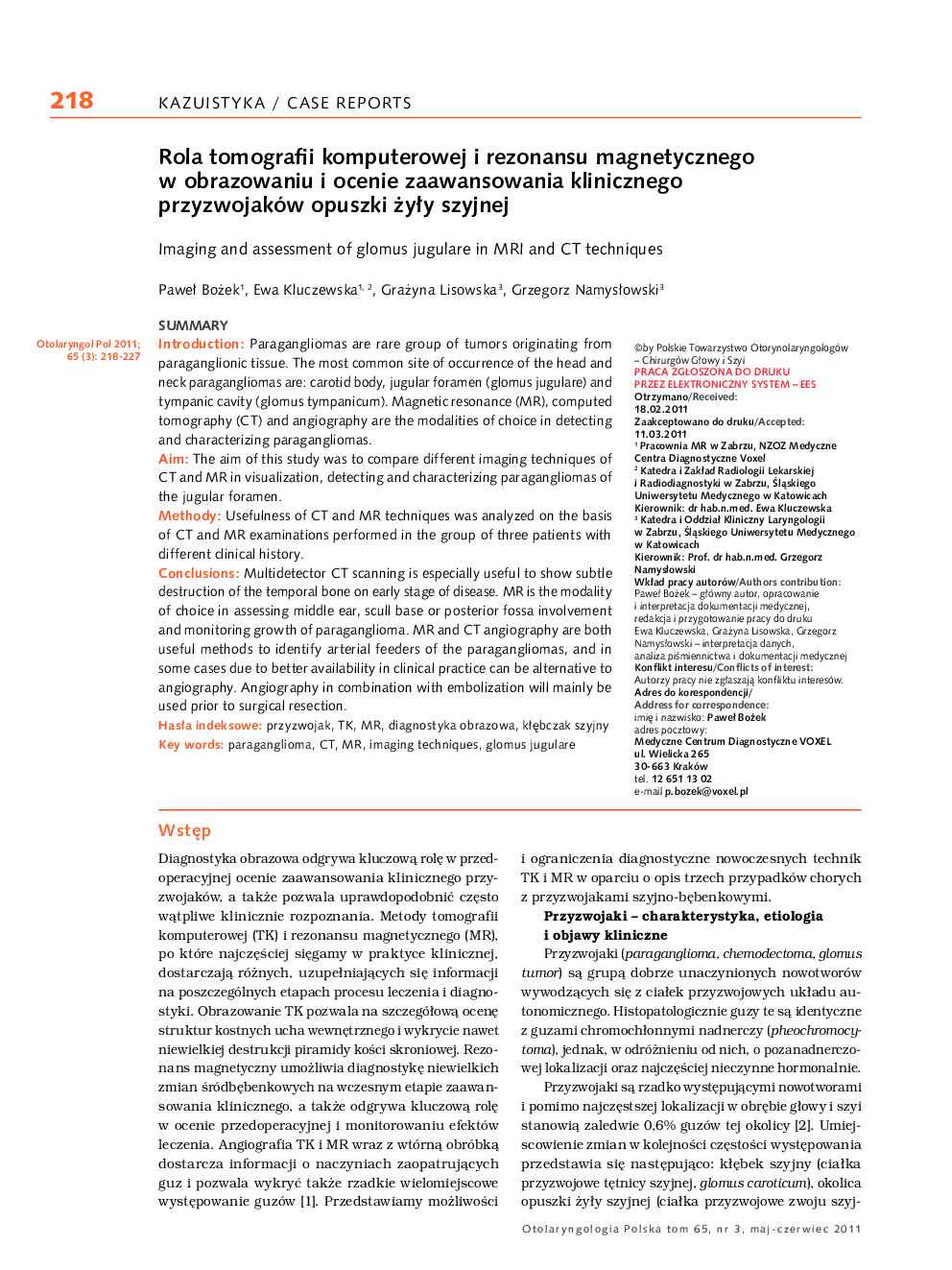
پیش نمایش صفحه اول مقاله

چکیده انگلیسی
Multidetector CT scanning is especially useful to show subtle destruction of the temporal bone on early stage of disease. MR is the modality of choice in assessing middle ear, scull base or posterior fossa involvement and monitoring growth of paraganglioma. MR and CT angiography are both useful methods to identify arterial feeders of the paragangliomas, and in some cases due to better availability in clinical practice can be alternative to angiography. Angiography in combination with embolization will mainly be used prior to surgical resection.
ناشر
Database: Elsevier - ScienceDirect (ساینس دایرکت)
Journal: Otolaryngologia Polska - Volume 65, Issue 3, MayâJune 2011, Pages 218-227
Journal: Otolaryngologia Polska - Volume 65, Issue 3, MayâJune 2011, Pages 218-227
نویسندگان
PaweÅ Bożek, Ewa Kluczewska, Grażyna Lisowska, Grzegorz NamysÅowski,