| کد مقاله | کد نشریه | سال انتشار | مقاله انگلیسی | نسخه تمام متن |
|---|---|---|---|---|
| 3271087 | 1208259 | 2010 | 8 صفحه PDF | دانلود رایگان |
عنوان انگلیسی مقاله ISI
Comparison of pQCT-Based Measures of Radial Bone Geometry and Apparent Trabecular Bone Structure Using Manufacturer and In-House-Developed Algorithms
دانلود مقاله + سفارش ترجمه
دانلود مقاله ISI انگلیسی
رایگان برای ایرانیان
کلمات کلیدی
موضوعات مرتبط
علوم پزشکی و سلامت
پزشکی و دندانپزشکی
غدد درون ریز، دیابت و متابولیسم
پیش نمایش صفحه اول مقاله

چکیده انگلیسی
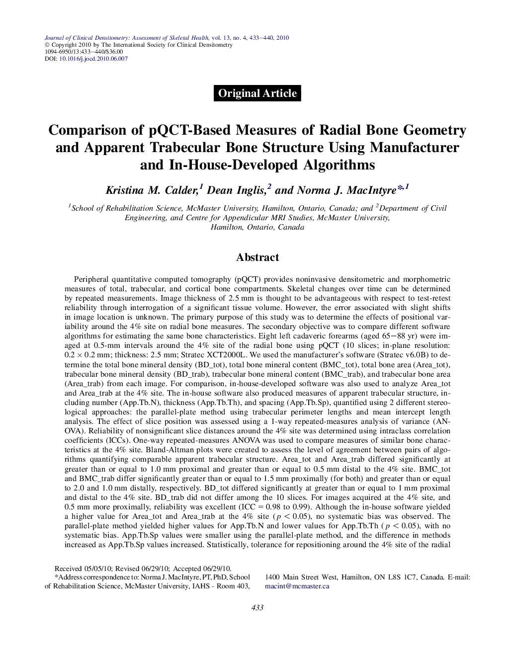
Peripheral quantitative computed tomography (pQCT) provides noninvasive densitometric and morphometric measures of total, trabecular, and cortical bone compartments. Skeletal changes over time can be determined by repeated measurements. Image thickness of 2.5 mm is thought to be advantageous with respect to test-retest reliability through interrogation of a significant tissue volume. However, the error associated with slight shifts in image location is unknown. The primary purpose of this study was to determine the effects of positional variability around the 4% site on radial bone measures. The secondary objective was to compare different software algorithms for estimating the same bone characteristics. Eight left cadaveric forearms (aged 65-88 yr) were imaged at 0.5-mm intervals around the 4% site of the radial bone using pQCT (10 slices; in-plane resolution: 0.2 Ã 0.2 mm; thickness: 2.5 mm; Stratec XCT2000L. We used the manufacturer's software (Stratec v6.0B) to determine the total bone mineral density (BD_tot), total bone mineral content (BMC_tot), total bone area (Area_tot), trabecular bone mineral density (BD_trab), trabecular bone mineral content (BMC_trab), and trabecular bone area (Area_trab) from each image. For comparison, in-house-developed software was also used to analyze Area_tot and Area_trab at the 4% site. The in-house software also produced measures of apparent trabecular structure, including number (App.Tb.N), thickness (App.Tb.Th), and spacing (App.Tb.Sp), quantified using 2 different stereological approaches: the parallel-plate method using trabecular perimeter lengths and mean intercept length analysis. The effect of slice position was assessed using a 1-way repeated-measures analysis of variance (ANOVA). Reliability of nonsignificant slice distances around the 4% site was determined using intraclass correlation coefficients (ICCs). One-way repeated-measures ANOVA was used to compare measures of similar bone characteristics at the 4% site. Bland-Altman plots were created to assess the level of agreement between pairs of algorithms quantifying comparable apparent trabecular structure. Area_tot and Area_trab differed significantly at greater than or equal to 1.0 mm proximal and greater than or equal to 0.5 mm distal to the 4% site. BMC_tot and BMC_trab differ significantly greater than or equal to 1.5 mm proximally (for both) and greater than or equal to 2.0 and 1.0 mm distally, respectively. BD_tot differed significantly at greater than or equal to 1 mm proximal and distal to the 4% site. BD_trab did not differ among the 10 slices. For images acquired at the 4% site, and 0.5 mm more proximally, reliability was excellent (ICC = 0.98 to 0.99). Although the in-house software yielded a higher value for Area_tot and Area_trab at the 4% site (p < 0.05), no systematic bias was observed. The parallel-plate method yielded higher values for App.Tb.N and lower values for App.Tb.Th (p < 0.05), with no systematic bias. App.Tb.Sp values were smaller using the parallel-plate method, and the difference in methods increased as App.Tb.Sp values increased. Statistically, tolerance for repositioning around the 4% site of the radial bone is least for measures of bone area and greatest for BD_trab. On repeated measures, a proximal shift of 0.5 mm will not influence the results.
ناشر
Database: Elsevier - ScienceDirect (ساینس دایرکت)
Journal: Journal of Clinical Densitometry - Volume 13, Issue 4, October 2010, Pages 433-440
Journal: Journal of Clinical Densitometry - Volume 13, Issue 4, October 2010, Pages 433-440
نویسندگان
Kristina M. Calder, Dean Inglis, Norma J. MacIntyre,