| کد مقاله | کد نشریه | سال انتشار | مقاله انگلیسی | نسخه تمام متن |
|---|---|---|---|---|
| 3271115 | 1208261 | 2012 | 11 صفحه PDF | دانلود رایگان |
عنوان انگلیسی مقاله ISI
Comparison Between Amputation-Induced Demineralization and Age-Related Bone Loss Using Digital X-Ray Radiogrammetry
دانلود مقاله + سفارش ترجمه
دانلود مقاله ISI انگلیسی
رایگان برای ایرانیان
کلمات کلیدی
موضوعات مرتبط
علوم پزشکی و سلامت
پزشکی و دندانپزشکی
غدد درون ریز، دیابت و متابولیسم
پیش نمایش صفحه اول مقاله

چکیده انگلیسی
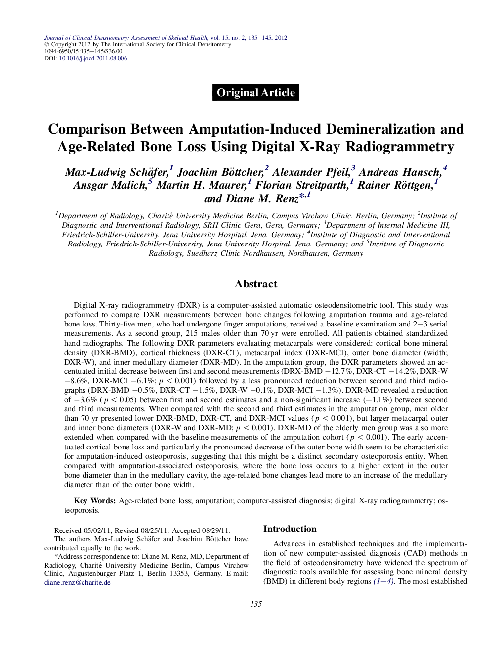
Digital X-ray radiogrammetry (DXR) is a computer-assisted automatic osteodensitometric tool. This study was performed to compare DXR measurements between bone changes following amputation trauma and age-related bone loss. Thirty-five men, who had undergone finger amputations, received a baseline examination and 2-3 serial measurements. As a second group, 215 males older than 70 yr were enrolled. All patients obtained standardized hand radiographs. The following DXR parameters evaluating metacarpals were considered: cortical bone mineral density (DXR-BMD), cortical thickness (DXR-CT), metacarpal index (DXR-MCI), outer bone diameter (width; DXR-W), and inner medullary diameter (DXR-MD). In the amputation group, the DXR parameters showed an accentuated initial decrease between first and second measurements (DRX-BMD â12.7%, DXR-CT â14.2%, DXR-W â8.6%, DXR-MCI â6.1%; p < 0.001) followed by a less pronounced reduction between second and third radiographs (DRX-BMD â0.5%, DXR-CT â1.5%, DXR-W â0.1%, DXR-MCI â1.3%). DXR-MD revealed a reduction of â3.6% (p < 0.05) between first and second estimates and a non-significant increase (+1.1%) between second and third measurements. When compared with the second and third estimates in the amputation group, men older than 70 yr presented lower DXR-BMD, DXR-CT, and DXR-MCI values (p < 0.001), but larger metacarpal outer and inner bone diameters (DXR-W and DXR-MD; p < 0.001). DXR-MD of the elderly men group was also more extended when compared with the baseline measurements of the amputation cohort (p < 0.001). The early accentuated cortical bone loss and particularly the pronounced decrease of the outer bone width seem to be characteristic for amputation-induced osteoporosis, suggesting that this might be a distinct secondary osteoporosis entity. When compared with amputation-associated osteoporosis, where the bone loss occurs to a higher extent in the outer bone diameter than in the medullary cavity, the age-related bone changes lead more to an increase of the medullary diameter than of the outer bone width.
ناشر
Database: Elsevier - ScienceDirect (ساینس دایرکت)
Journal: Journal of Clinical Densitometry - Volume 15, Issue 2, AprilâJune 2012, Pages 135-145
Journal: Journal of Clinical Densitometry - Volume 15, Issue 2, AprilâJune 2012, Pages 135-145
نویسندگان
Max-Ludwig Schäfer, Joachim Böttcher, Alexander Pfeil, Andreas Hansch, Ansgar Malich, Martin H. Maurer, Florian Streitparth, Rainer Röttgen, Diane M. Renz,