| کد مقاله | کد نشریه | سال انتشار | مقاله انگلیسی | نسخه تمام متن |
|---|---|---|---|---|
| 3271980 | 1208289 | 2006 | 6 صفحه PDF | دانلود رایگان |
عنوان انگلیسی مقاله ISI
Assessment of Femoral Neck Strength by 3-Dimensional X-ray Absorptiometry
دانلود مقاله + سفارش ترجمه
دانلود مقاله ISI انگلیسی
رایگان برای ایرانیان
موضوعات مرتبط
علوم پزشکی و سلامت
پزشکی و دندانپزشکی
غدد درون ریز، دیابت و متابولیسم
پیش نمایش صفحه اول مقاله

چکیده انگلیسی
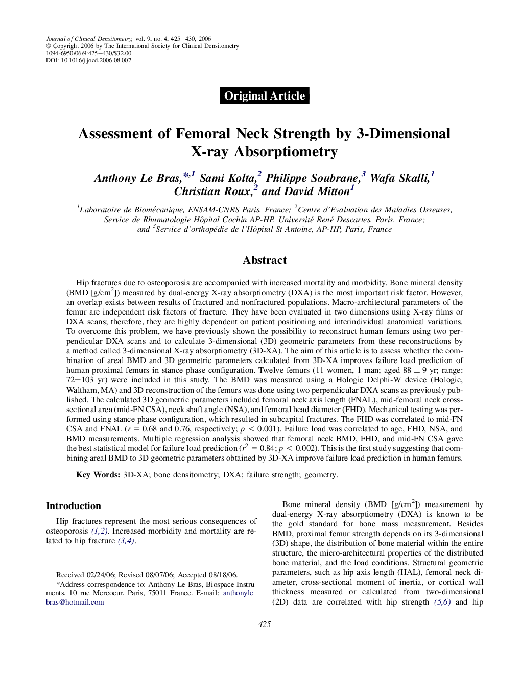
Hip fractures due to osteoporosis are accompanied with increased mortality and morbidity. Bone mineral density (BMD [g/cm2]) measured by dual-energy X-ray absorptiometry (DXA) is the most important risk factor. However, an overlap exists between results of fractured and nonfractured populations. Macro-architectural parameters of the femur are independent risk factors of fracture. They have been evaluated in two dimensions using X-ray films or DXA scans; therefore, they are highly dependent on patient positioning and interindividual anatomical variations. To overcome this problem, we have previously shown the possibility to reconstruct human femurs using two perpendicular DXA scans and to calculate 3-dimensional (3D) geometric parameters from these reconstructions by a method called 3-dimensional X-ray absorptiometry (3D-XA). The aim of this article is to assess whether the combination of areal BMD and 3D geometric parameters calculated from 3D-XA improves failure load prediction of human proximal femurs in stance phase configuration. Twelve femurs (11 women, 1 man; aged 88 ± 9 yr; range: 72-103 yr) were included in this study. The BMD was measured using a Hologic Delphi-W device (Hologic, Waltham, MA) and 3D reconstruction of the femurs was done using two perpendicular DXA scans as previously published. The calculated 3D geometric parameters included femoral neck axis length (FNAL), mid-femoral neck cross-sectional area (mid-FN CSA), neck shaft angle (NSA), and femoral head diameter (FHD). Mechanical testing was performed using stance phase configuration, which resulted in subcapital fractures. The FHD was correlated to mid-FN CSA and FNAL (r = 0.68 and 0.76, respectively; p < 0.001). Failure load was correlated to age, FHD, NSA, and BMD measurements. Multiple regression analysis showed that femoral neck BMD, FHD, and mid-FN CSA gave the best statistical model for failure load prediction (r2 = 0.84; p < 0.002). This is the first study suggesting that combining areal BMD to 3D geometric parameters obtained by 3D-XA improve failure load prediction in human femurs.
ناشر
Database: Elsevier - ScienceDirect (ساینس دایرکت)
Journal: Journal of Clinical Densitometry - Volume 9, Issue 4, OctoberâDecember 2006, Pages 425-430
Journal: Journal of Clinical Densitometry - Volume 9, Issue 4, OctoberâDecember 2006, Pages 425-430
نویسندگان
Anthony Le Bras, Sami Kolta, Philippe Soubrane, Wafa Skalli, Christian Roux, David Mitton,