| کد مقاله | کد نشریه | سال انتشار | مقاله انگلیسی | نسخه تمام متن |
|---|---|---|---|---|
| 3979062 | 1257317 | 2012 | 12 صفحه PDF | دانلود رایگان |
عنوان انگلیسی مقاله ISI
Imagerie rénale : anatomie radiologique et méthodes d'exploration
دانلود مقاله + سفارش ترجمه
دانلود مقاله ISI انگلیسی
رایگان برای ایرانیان
کلمات کلیدی
IRMÉchographie - اسکنScanner - اسکنرMRI - امآرآی یا تصویرسازی تشدید مغناطیسیMultimodality imaging - تصویربرداری چندمتغیریToxicité - سمیتScintigraphie - سینتی گرافیScintigraphy - سینتیگرافی، درخشه نگاریContrast agent - عامل کنتراستUltrasound - فراصوتProduit de Contraste - محصول کنتراستCT scan - مقطعنگاری رایانهای یا برشنگاری رایانهای یا توموگرافی رایانهایBilan d’extension - گزارش فرمت
موضوعات مرتبط
علوم پزشکی و سلامت
پزشکی و دندانپزشکی
تومور شناسی
پیش نمایش صفحه اول مقاله

چکیده انگلیسی
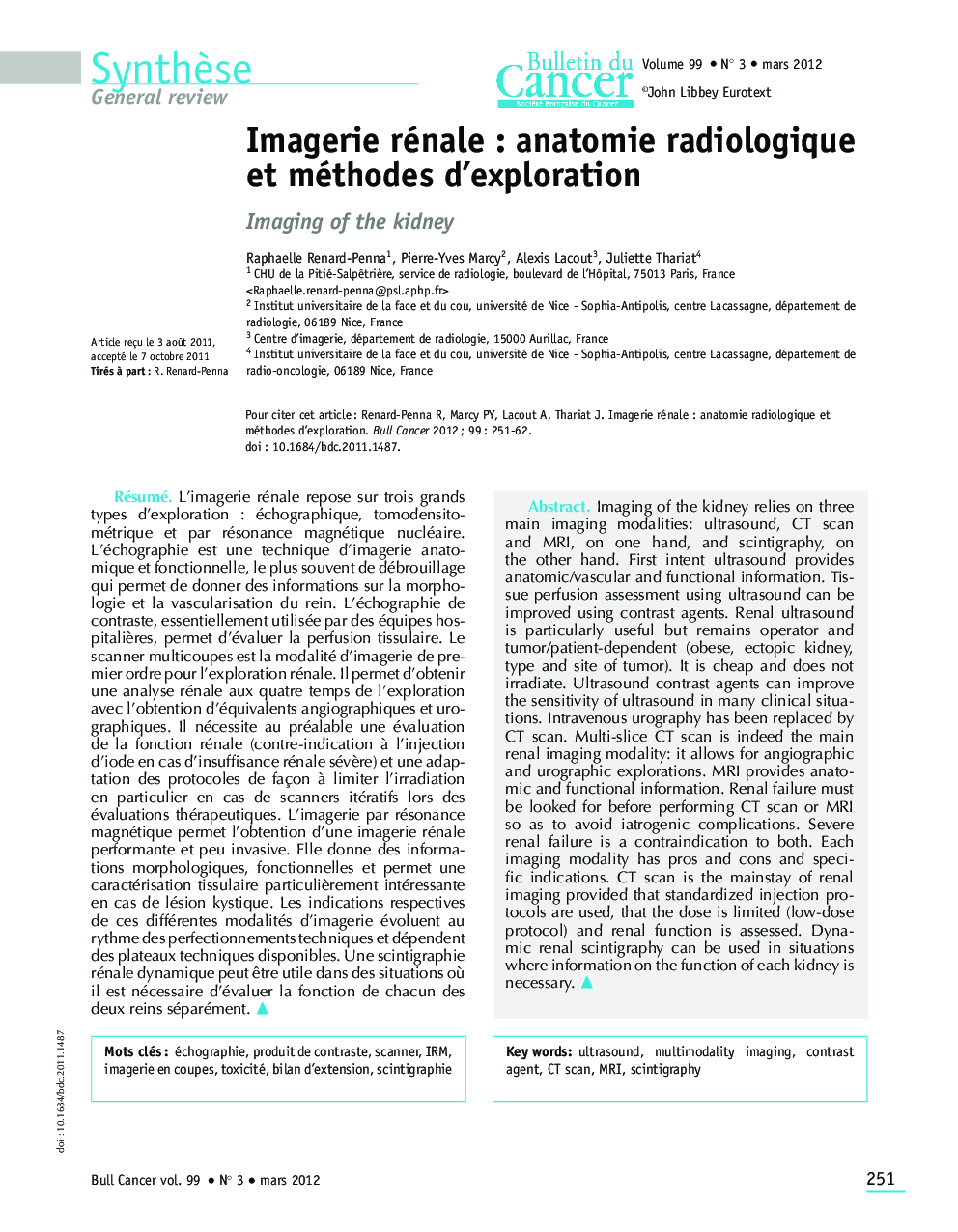
Imaging of the kidney relies on three main imaging modalities: ultrasound, CT scan and MRI, on one hand, and scintigraphy, on the other hand. First intent ultrasound provides anatomic/vascular and functional information. Tissue perfusion assessment using ultrasound can be improved using contrast agents. Renal ultrasound is particularly useful but remains operator and tumor/patient-dependent (obese, ectopic kidney, type and site of tumor). It is cheap and does not irradiate. Ultrasound contrast agents can improve the sensitivity of ultrasound in many clinical situations. Intravenous urography has been replaced by CT scan. Multi-slice CT scan is indeed the main renal imaging modality: it allows for angiographic and urographic explorations. MRI provides anatomic and functional information. Renal failure must be looked for before performing CT scan or MRI so as to avoid iatrogenic complications. Severe renal failure is a contraindication to both. Each imaging modality has pros and cons and specific indications. CT scan is the mainstay of renal imaging provided that standardized injection protocols are used, that the dose is limited (low-dose protocol) and renal function is assessed. Dynamic renal scintigraphy can be used in situations where information on the function of each kidney is necessary.
ناشر
Database: Elsevier - ScienceDirect (ساینس دایرکت)
Journal: Bulletin du Cancer - Volume 99, Issue 3, March 2012, Pages 251-262
Journal: Bulletin du Cancer - Volume 99, Issue 3, March 2012, Pages 251-262
نویسندگان
Raphaelle Renard-Penna, Pierre-Yves Marcy, Alexis Lacout, Juliette Thariat,