| کد مقاله | کد نشریه | سال انتشار | مقاله انگلیسی | نسخه تمام متن |
|---|---|---|---|---|
| 3982496 | 1257729 | 2013 | 6 صفحه PDF | دانلود رایگان |
عنوان انگلیسی مقاله ISI
Intercostal lung hernia: Radiographic and MDCT findings
دانلود مقاله + سفارش ترجمه
دانلود مقاله ISI انگلیسی
رایگان برای ایرانیان
موضوعات مرتبط
علوم پزشکی و سلامت
پزشکی و دندانپزشکی
تومور شناسی
پیش نمایش صفحه اول مقاله

چکیده انگلیسی
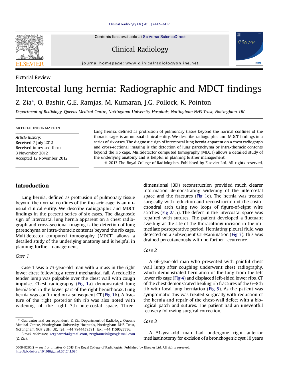
Lung hernia, defined as protrusion of pulmonary tissue beyond the normal confines of the thoracic cage, is an unusual clinical entity. We describe radiographic and MDCT findings in a series of six cases. The diagnostic sign of intercostal lung hernia apparent on a chest radiograph and cross-sectional imaging is the detection of lung parenchyma or intra-thoracic contents beyond the rib cage. Multidetector computed tomography (MDCT) allows a detailed study of the underlying anatomy and is helpful in planning further management.
ناشر
Database: Elsevier - ScienceDirect (ساینس دایرکت)
Journal: Clinical Radiology - Volume 68, Issue 7, July 2013, Pages e412–e417
Journal: Clinical Radiology - Volume 68, Issue 7, July 2013, Pages e412–e417
نویسندگان
Z. Zia, O. Bashir, G.E. Ramjas, M. Kumaran, J.G. Pollock, K. Pointon,