| کد مقاله | کد نشریه | سال انتشار | مقاله انگلیسی | نسخه تمام متن |
|---|---|---|---|---|
| 3983316 | 1257760 | 2009 | 10 صفحه PDF | دانلود رایگان |
عنوان انگلیسی مقاله ISI
Imaging in pulsatile tinnitus
دانلود مقاله + سفارش ترجمه
دانلود مقاله ISI انگلیسی
رایگان برای ایرانیان
موضوعات مرتبط
علوم پزشکی و سلامت
پزشکی و دندانپزشکی
تومور شناسی
پیش نمایش صفحه اول مقاله

چکیده انگلیسی
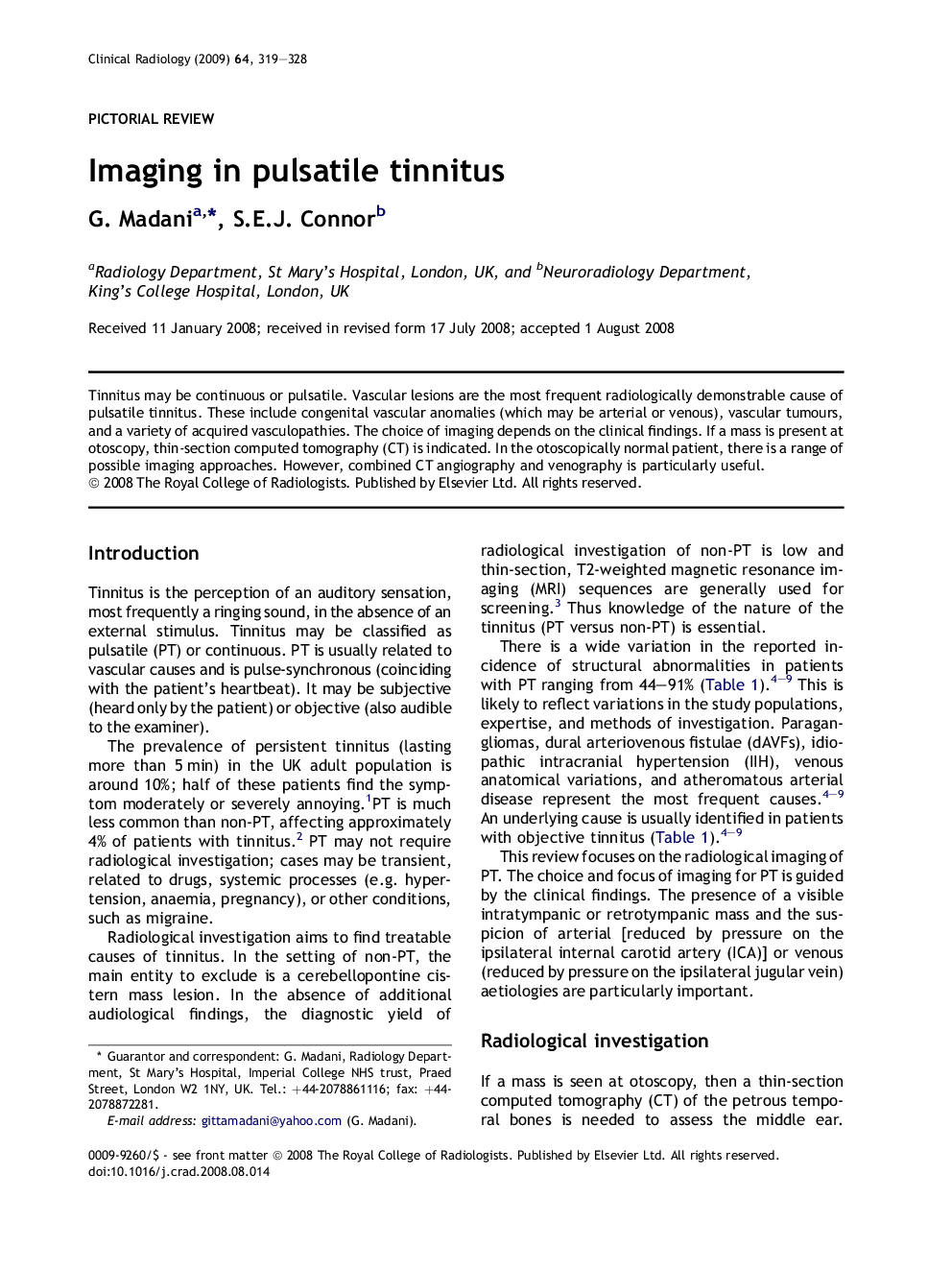
Tinnitus may be continuous or pulsatile. Vascular lesions are the most frequent radiologically demonstrable cause of pulsatile tinnitus. These include congenital vascular anomalies (which may be arterial or venous), vascular tumours, and a variety of acquired vasculopathies. The choice of imaging depends on the clinical findings. If a mass is present at otoscopy, thin-section computed tomography (CT) is indicated. In the otoscopically normal patient, there is a range of possible imaging approaches. However, combined CT angiography and venography is particularly useful.
ناشر
Database: Elsevier - ScienceDirect (ساینس دایرکت)
Journal: Clinical Radiology - Volume 64, Issue 3, March 2009, Pages 319–328
Journal: Clinical Radiology - Volume 64, Issue 3, March 2009, Pages 319–328
نویسندگان
G. Madani, S.E.J. Connor,