| کد مقاله | کد نشریه | سال انتشار | مقاله انگلیسی | نسخه تمام متن |
|---|---|---|---|---|
| 4227944 | 1609833 | 2008 | 5 صفحه PDF | دانلود رایگان |
عنوان انگلیسی مقاله ISI
Monochromatic beam characterization for Auger electron dosimetry and radiotherapy
دانلود مقاله + سفارش ترجمه
دانلود مقاله ISI انگلیسی
رایگان برای ایرانیان
موضوعات مرتبط
علوم پزشکی و سلامت
پزشکی و دندانپزشکی
رادیولوژی و تصویربرداری
پیش نمایش صفحه اول مقاله

چکیده انگلیسی
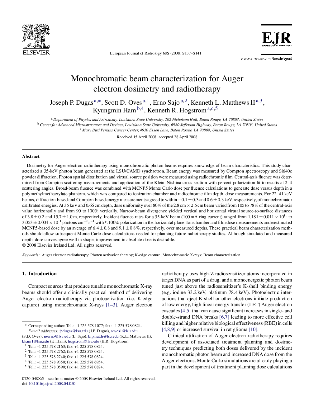
Dosimetry for Auger electron radiotherapy using monochromatic photon beams requires knowledge of beam characteristics. This study characterized a 35-keV photon beam generated at the LSU/CAMD synchrotron. Beam energy was measured by Compton spectroscopy and Si640c powder diffraction. Photon spatial distribution and virtual source position were measured using radiochromic film. Central-axis fluence was determined from Compton scattering measurements and application of the Klein-Nishina cross-section with percent polarization fit to results at 2-4 scattering angles. Broad-beam fluence was combined with MCNP5 Monte Carlo dose per fluence calculations to generate dose versus depth in a polymethylmethacrylate phantom, which was compared to ionization chamber and radiochromic film depth-dose measurements. For 22-41 keV beams, diffraction-based and Compton-based energy measurements agreed to within â0.1 ± 0.3 and 0.6 ± 0.3 keV, respectively, of monochromator calibrated energies. At 35 keV and 0.66 cm depth, dose uniformity over 80% of the 2.8 cm Ã 2.5 cm beam varied from 105 to 78% of the central-axis value horizontally and from 90 to 100% vertically. Narrow-beam divergence yielded vertical and horizontal virtual source-to-surface distances of 3.8 ± 0.2 and 15.7 ± 1.0 m, respectively. Incident fluence rates for a 35-keV beam (100 mA ring current) ranged from 1.181 ± 0.011 Ã 1011 to 3.053 ± 0.004 Ã 1011 photons cmâ2 sâ1 with â100% polarization in the horizontal plane. Ion chamber and film dose measurements underestimated MCNP5-based dose by an average of 6.4 ± 0.8 and 9.1 ± 0.8%, respectively, over measured depths. These practical beam characterization methods should allow subsequent Monte Carlo dose calculations needed for planning future radiotherapy studies. Although simulated and measured depth-dose curves agree well in shape, improvement in absolute dose is desirable.
ناشر
Database: Elsevier - ScienceDirect (ساینس دایرکت)
Journal: European Journal of Radiology - Volume 68, Issue 3, Supplement, December 2008, Pages S137-S141
Journal: European Journal of Radiology - Volume 68, Issue 3, Supplement, December 2008, Pages S137-S141
نویسندگان
Joseph P. Dugas, Scott D. Oves, Erno Sajo, Kenneth L. II, Kyungmin Ham, Kenneth R. Hogstrom,