| کد مقاله | کد نشریه | سال انتشار | مقاله انگلیسی | نسخه تمام متن |
|---|---|---|---|---|
| 592391 | 1453904 | 2015 | 7 صفحه PDF | دانلود رایگان |
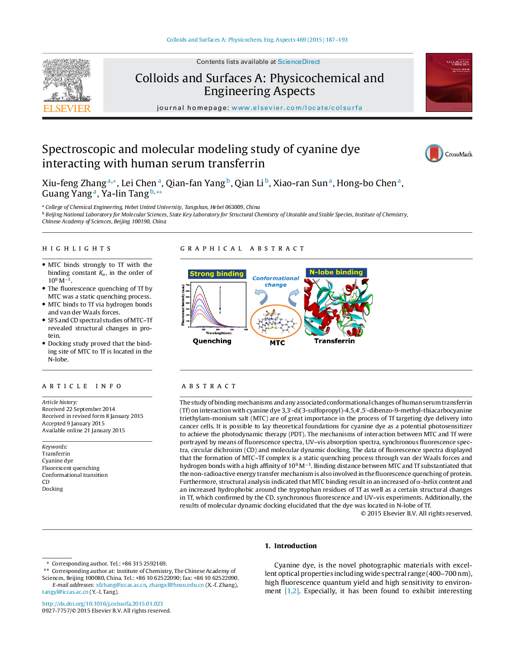
• MTC binds strongly to Tf with the binding constant Ka, in the order of 109 M−1.
• The fluorescence quenching of Tf by MTC was a static quenching process.
• MTC binds to Tf via hydrogen bonds and van der Waals forces.
• SFS and CD spectral studies of MTC–Tf revealed structural changes in protein.
• Docking study proved that the binding site of MTC to Tf is located in the N-lobe.
The study of binding mechanisms and any associated conformational changes of human serum transferrin (Tf) on interaction with cyanine dye 3,3′-di(3-sulfopropyl)-4,5,4′,5′-dibenzo-9-methyl-thiacarbocyanine triethylam-monium salt (MTC) are of great importance in the process of Tf targeting dye delivery into cancer cells. It is possible to lay theoretical foundations for cyanine dye as a potential photosensitizer to achieve the photodynamic therapy (PDT). The mechanisms of interaction between MTC and Tf were portrayed by means of fluorescence spectra, UV–vis absorption spectra, synchronous fluorescence spectra, circular dichroism (CD) and molecular dynamic docking. The data of fluorescence spectra displayed that the formation of MTC–Tf complex is a static quenching process through van der Waals forces and hydrogen bonds with a high affinity of 109 M−1. Binding distance between MTC and Tf substantiated that the non-radioactive energy transfer mechanism is also involved in the fluorescence quenching of protein. Furthermore, structural analysis indicated that MTC binding result in an increased of α-helix content and an increased hydrophobic around the tryptophan residues of Tf as well as a certain structural changes in Tf, which confirmed by the CD, synchronous fluorescence and UV–vis experiments. Additionally, the results of molecular dynamic docking elucidated that the dye was located in N-lobe of Tf.
Figure optionsDownload as PowerPoint slide
Journal: Colloids and Surfaces A: Physicochemical and Engineering Aspects - Volume 469, 20 March 2015, Pages 187–193
