| کد مقاله | کد نشریه | سال انتشار | مقاله انگلیسی | نسخه تمام متن |
|---|---|---|---|---|
| 5980310 | 1176913 | 2014 | 11 صفحه PDF | دانلود رایگان |

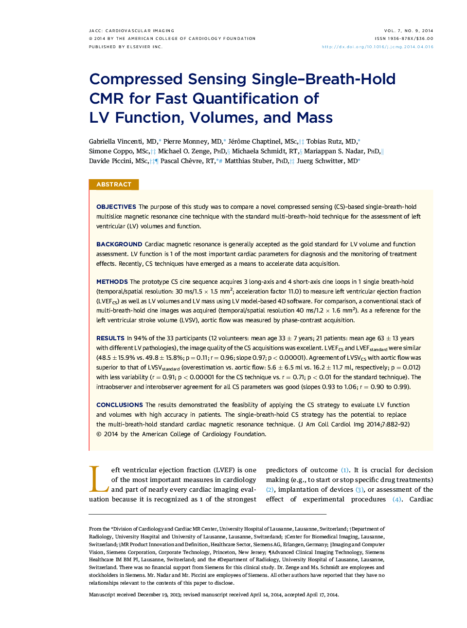
ObjectivesThe purpose of this study was to compare a novel compressed sensing (CS)-based single-breath-hold multislice magnetic resonance cine technique with the standard multi-breath-hold technique for the assessment of left ventricular (LV) volumes and function.BackgroundCardiac magnetic resonance is generally accepted as the gold standard for LV volume and function assessment. LV function is 1 of the most important cardiac parameters for diagnosis and the monitoring of treatment effects. Recently, CS techniques have emerged as a means to accelerate data acquisition.MethodsThe prototype CS cine sequence acquires 3 long-axis and 4 short-axis cine loops in 1 single breath-hold (temporal/spatial resolution: 30 ms/1.5 à 1.5 mm2; acceleration factor 11.0) to measure left ventricular ejection fraction (LVEFCS) as well as LV volumes and LV mass using LV model-based 4D software. For comparison, a conventional stack of multi-breath-hold cine images was acquired (temporal/spatial resolution 40 ms/1.2 à 1.6 mm2). As a reference for the left ventricular stroke volume (LVSV), aortic flow was measured by phase-contrast acquisition.ResultsIn 94% of the 33 participants (12 volunteers: mean age 33 ± 7 years; 21 patients: mean age 63 ± 13 years with different LV pathologies), the image quality of the CS acquisitions was excellent. LVEFCS and LVEFstandard were similar (48.5 ± 15.9% vs. 49.8 ± 15.8%; p = 0.11; r = 0.96; slope 0.97; p < 0.00001). Agreement of LVSVCS with aortic flow was superior to that of LVSVstandard (overestimation vs. aortic flow: 5.6 ± 6.5 ml vs. 16.2 ± 11.7 ml, respectively; p = 0.012) with less variability (r = 0.91; p < 0.00001 for the CS technique vs. r = 0.71; p < 0.01 for the standard technique). The intraobserver and interobserver agreement for all CS parameters was good (slopes 0.93 to 1.06; r = 0.90 to 0.99).ConclusionsThe results demonstrated the feasibility of applying the CS strategy to evaluate LV function and volumes with high accuracy in patients. The single-breath-hold CS strategy has the potential to replace the multi-breath-hold standard cardiac magnetic resonance technique.
Journal: JACC: Cardiovascular Imaging - Volume 7, Issue 9, September 2014, Pages 882-892