| کد مقاله | کد نشریه | سال انتشار | مقاله انگلیسی | نسخه تمام متن |
|---|---|---|---|---|
| 9390227 | 1282787 | 2005 | 4 صفحه PDF | دانلود رایگان |
عنوان انگلیسی مقاله ISI
Tumeur mélanocytaire méningée primitive de grade intermédiaire : confrontation radio-pathologique
دانلود مقاله + سفارش ترجمه
دانلود مقاله ISI انگلیسی
رایگان برای ایرانیان
کلمات کلیدی
موضوعات مرتبط
علوم پزشکی و سلامت
پزشکی و دندانپزشکی
رادیولوژی و تصویربرداری
پیش نمایش صفحه اول مقاله

چکیده انگلیسی
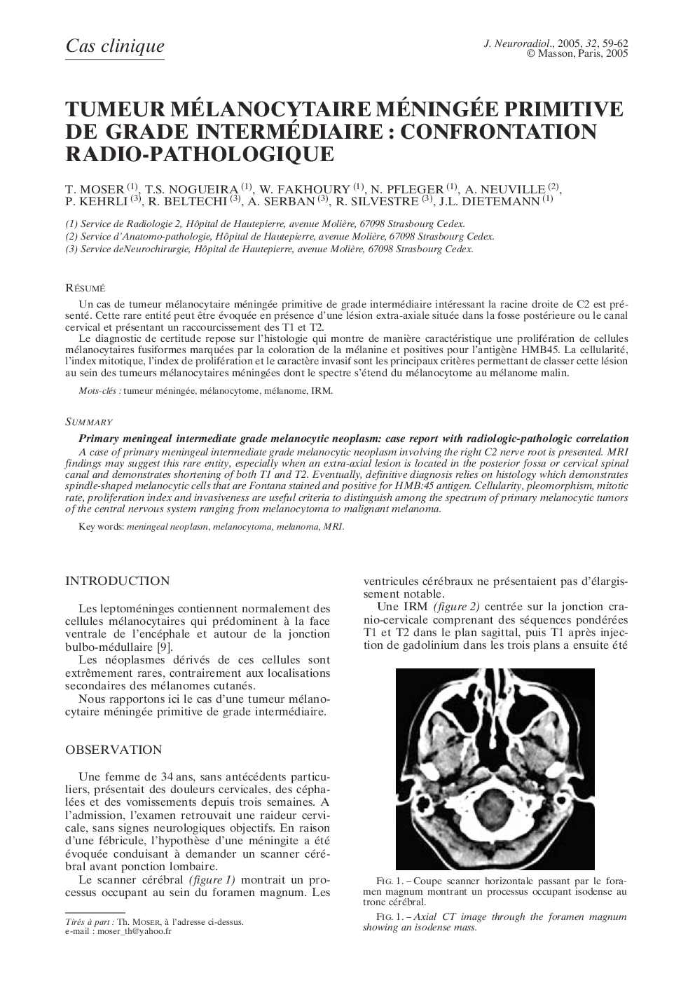
A case of primary meningeal intermediate grade melanocytic neoplasm involving the right C2 nerve root is presented. MRI findings may suggest this rare entity, especially when an extra-axial lesion is located in the posterior fossa or cervical spinal canal and demonstrates shortening of both T1 and T2. Eventually, definitive diagnosis relies on histology which demonstrates spindle-shaped melanocytic cells that are Fontana stained and positive for HMB:45 antigen. Cellularity, pleomorphism, mitotic rate, proliferation index and invasiveness are useful criteria to distinguish among the spectrum of primary melanocytic tumors of the central nervous system ranging from melanocytoma to malignant melanoma.
ناشر
Database: Elsevier - ScienceDirect (ساینس دایرکت)
Journal: Journal of Neuroradiology - Volume 32, Issue 1, January 2005, Pages 59-62
Journal: Journal of Neuroradiology - Volume 32, Issue 1, January 2005, Pages 59-62
نویسندگان
T. Moser, T.S. Nogueira, W. Fakhoury, N. Pfleger, A. Neuville, P. Kehrli, R. Beltechi, A. Serban, R. Silvestre, J.L. Dietemann,