| کد مقاله | کد نشریه | سال انتشار | مقاله انگلیسی | نسخه تمام متن |
|---|---|---|---|---|
| 9394040 | 1283968 | 2005 | 11 صفحه PDF | دانلود رایگان |
عنوان انگلیسی مقاله ISI
Evaluation of Therapy for Lymphoma
دانلود مقاله + سفارش ترجمه
دانلود مقاله ISI انگلیسی
رایگان برای ایرانیان
موضوعات مرتبط
علوم پزشکی و سلامت
پزشکی و دندانپزشکی
رادیولوژی و تصویربرداری
پیش نمایش صفحه اول مقاله

چکیده انگلیسی
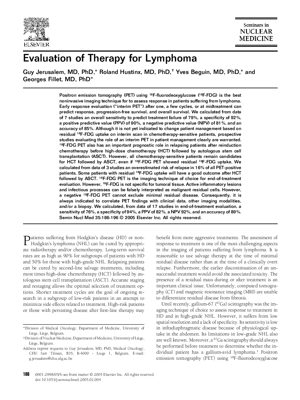
Positron emission tomography (PET) using 18F-fluorodeoxyglucose (18F-FDG) is the best noninvasive imaging technique for to assess response in patients suffering from lymphoma. Early response evaluation (“interim PET”) after one, a few cycles, or at midtreatment can predict response, progression-free survival, and overall survival. We calculated from data of 7 studies an overall sensitivity to predict treatment failure of 79%, a specificity of 92%, a positive predictive value (PPV) of 90%, a negative predictive value (NPV) of 81%, and an accuracy of 85%. Although it is not yet indicated to change patient management based on residual 18F-FDG uptake on interim scan in chemotherapy-sensitive patients, prospective studies evaluating the role of an interim PET in patient management clearly are warranted. 18F-FDG PET also has an important prognostic role in relapsing patients after reinduction chemotherapy before high-dose chemotherapy (HCT) followed by autologous stem cell transplantation (ASCT). However, all chemotherapy-sensitive patients remain candidates for HCT followed by ASCT, even if 18F-FDG PET showed residual 18F-FDG uptake. We calculated from data of 3 studies an overestimated risk of relapse in 16% of all PET-positive patients. Some patients with residual 18F-FDG uptake will have a good outcome after HCT followed by ASCT. 18F-FDG PET is the imaging technique of choice for end-of-treatment evaluation. However, 18F-FDG is not specific for tumoral tissue. Active inflammatory lesions and infectious processes can be falsely interpreted as malignant residual cells. However, a negative 18F-FDG PET cannot exclude minimal residual disease. Consequently, it is always indicated to correlate PET findings with clinical data, other imaging modalities, and/or a biopsy. We calculated, from data of 17 studies in end-of-treatment evaluation, a sensitivity of 76%, a specificity of 94%, a PPV of 82%, a NPV 92%, and an accuracy of 89%.
ناشر
Database: Elsevier - ScienceDirect (ساینس دایرکت)
Journal: Seminars in Nuclear Medicine - Volume 35, Issue 3, July 2005, Pages 186-196
Journal: Seminars in Nuclear Medicine - Volume 35, Issue 3, July 2005, Pages 186-196
نویسندگان
Guy MD, PhD, Roland MD, PhD, Yves MD, PhD, Georges MD, PhD,