| کد مقاله | کد نشریه | سال انتشار | مقاله انگلیسی | نسخه تمام متن |
|---|---|---|---|---|
| 10097613 | 1609880 | 2005 | 7 صفحه PDF | دانلود رایگان |
عنوان انگلیسی مقاله ISI
Comparison of quantitative and visual analysis of Tc-99m MIBI scintimammography for detection of primary breast cancer
دانلود مقاله + سفارش ترجمه
دانلود مقاله ISI انگلیسی
رایگان برای ایرانیان
کلمات کلیدی
موضوعات مرتبط
علوم پزشکی و سلامت
پزشکی و دندانپزشکی
رادیولوژی و تصویربرداری
پیش نمایش صفحه اول مقاله

چکیده انگلیسی
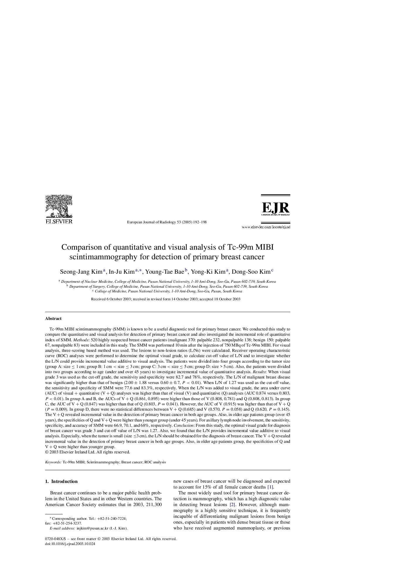
Tc-99m MIBI scintimammography (SMM) is known to be a useful diagnostic tool for primary breast cancer. We conducted this study to compare the quantitative and visual analysis for detection of primary breast cancer and also investigated the incremental role of quantitative index of SMM. Methods: 520 highly suspected breast cancer patients (malignant 370: palpable 232, nonpalpable 138; benign 150: palpable 67, nonpalpable 83) were included in this study. The SMM was performed 10 min after the injection of 750 MBq of Tc-99m MIBI. For visual analysis, three-scoring based method was used. The lesions to non-lesion ratios (L/Ns) were calculated. Receiver operating characteristic curve (ROC) analyses were performed to determine the optimal visual grade, to calculate cut-off value of L/N and to investigate whether the L/N could provide incremental value additive to visual analysis. The patients were divided into four groups according to the tumor size (group A: size ⤠1 cm; group B: 1 cm < size ⤠3 cm; group C: 3 cm < size ⤠5 cm; group D: size > 5 cm). Also, the patients were divided into two groups according to age (under and over 45 years) to investigate incremental value of quantitative analysis. Results: When visual grade 3 was used as the cut-off grade, the sensitivity and specificity were 82.7 and 78%, respectively. The L/N of malignant breast disease was significantly higher than that of benign (2.00±1.88 versus 0.60±0.7, P<0.01). When L/N of 1.27 was used as the cut-off value, the sensitivity and specificity of SMM were 77.6 and 83.3%, respectively. When the L/N was added to visual grade, the area under curve (AUC) of visual + quantitative (V+Q) analyses was higher than that of visual (V) and quantitative (Q) analyses (AUC 0.874 versus 0.803, P<0.01). In group A and B, the AUCs of V+Q (0.861, 0.895) were higher than those of V (0.808, 0.781) and Q (0.808, 0.813). In group C, the AUC of V+Q (0.847) was higher than that of Q (0.803, P=0.041). However, the AUC of V (0.915) was higher than that of V+Q (P=0.009). In group D, there were no statistical differences between V+Q (0.685) and V (0.570, P=0.058) and Q (0.620, P=0.145). The V+Q revealed incremental value in the detection of primary breast cancer in both age groups. Also, in older age patients group (over 45 years), the specificities of Q and V+Q were higher than younger group (under 45 years). For axillary lymph node involvement, the sensitivity, specificity, and accuracy of SMM were 66.9, 70.1, and 68%, respectively. Conclusion: From this study, the optimal visual grade for diagnosis of breast cancer was grade 3 and cut-off value of L/N was 1.27. Also, we found that the L/N provides incremental value additive to visual analysis. Especially, when the tumor is small (size â¤3 cm), the L/N should be obtained for the diagnosis of breast cancer. The V+Q revealed incremental value in the detection of primary breast cancer in both age groups. Also, in older age patients group, the specificities of Q and V+Q were higher than younger group.
ناشر
Database: Elsevier - ScienceDirect (ساینس دایرکت)
Journal: European Journal of Radiology - Volume 53, Issue 2, February 2005, Pages 192-198
Journal: European Journal of Radiology - Volume 53, Issue 2, February 2005, Pages 192-198
نویسندگان
Seong-Jang Kim, In-Ju Kim, Young-Tae Bae, Yong-Ki Kim, Dong-Soo Kim,