| کد مقاله | کد نشریه | سال انتشار | مقاله انگلیسی | نسخه تمام متن |
|---|---|---|---|---|
| 1995306 | 1064961 | 2009 | 20 صفحه PDF | دانلود رایگان |
عنوان انگلیسی مقاله ISI
PKCα-MAPK/ERK-phospholipase A2 signaling is required for human melanoma-enhanced brain endothelial cell proliferation and motility
دانلود مقاله + سفارش ترجمه
دانلود مقاله ISI انگلیسی
رایگان برای ایرانیان
کلمات کلیدی
COXiPLA2GAPDHCOX-2vWFERKVEGF-APKCcPLA2ERK1/2 - ERK1 / 2cyclooxygenase - آنزیم سیکلواکسیژنازtumor angiogenesis - آنژیوژنز تومورArachidonic acid - اسید آراشیدونیکEndothelial cell - سلول های اندوتلیالEndothelial cells - سلولهای اندوتلیالvascular endothelial growth factor-A - فاکتور رشد اندوتلیال عروق Acytosolic phospholipase A2 - فسفولیپاز A2 سیتوزولMelanomas - ملانوموسConfocal microscopy - میکروسکوپ کانفوکالprotein kinase C alpha - پروتئین کیناز C آلفاProtein kinase C - پروتئین کیناز سیmitogen activated protein kinase - پروتئین کیناز فعال Mitogen فعال استMAP kinase - کیناز MAPextracellular signal-regulated kinase - کیناز تنظیم شده سیگنال خارج سلولیglyceraldehyde-3-phosphate dehydrogenase - گلیسرالیدید-3-فسفات دهیدروژناز
موضوعات مرتبط
علوم زیستی و بیوفناوری
بیوشیمی، ژنتیک و زیست شناسی مولکولی
زیست شیمی
پیش نمایش صفحه اول مقاله

چکیده انگلیسی
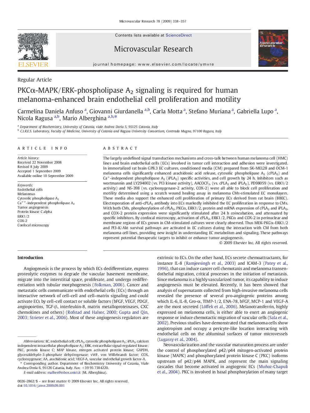
The largely undefined signal transduction mechanisms and cross-talk between human melanoma cell (HMC) lines and brain endothelial cells (ECs) involved in tumor cell interaction and adhesion were investigated. In immortalized rat brain GP8.3 EC cultures, conditioned media (CM) prepared from SK-MEL28 and OCM-1 melanoma cells significantly enhanced arachidonic acid release, cytosolic phospholipase A2 (cPLA2) and Ca+-independent phospholipase A2 (iPLA2) specific activities, and cell growth by 24 h. Inhibitors such as wortmannin and LY294002 (vs. PI3 kinase activity), AACOCF3, (vs. cPLA2 and iPLA2), PD98059 (vs. ERK1/2 activity) and NS-398 (vs. cyclooxygenase-2 activity, COX-2) were all able to block cell proliferation and motility determined using a scratch wound healing assay in melanoma CMs-stimulated EC monolayers. These media also support the enhanced cell proliferation of primary ECs derived from rat brain (BBEC). Electroporation of anti-cPLA2 antibody into ECs markedly inhibited the EC proliferation in response to CMs. With both CMs, phosphorylation of cPLA2, PKCα, ERK1/2, protein and mRNA expression of cPLA2 and iPLA2, and COX-2 protein expression were significantly stimulated after 24 h coincubation, and attenuated by specific inhibitors. By confocal microscopy, activation of cPLA2, ERK1/2, PKCα and COX-2 in perinuclear and membrane regions of ECs grown in CM-stimulated cultures were clearly observed. Thus MEK-PKCα-ERK1/2 and PI3-K/Akt survival pathways are activated in EC cultures during the interaction with CM from both melanoma cell lines, providing new insight in understanding EC metabolism and signaling. These pathways represent potential therapeutic targets to inhibit or enhance tumor angiogenesis.
ناشر
Database: Elsevier - ScienceDirect (ساینس دایرکت)
Journal: Microvascular Research - Volume 78, Issue 3, December 2009, Pages 338-357
Journal: Microvascular Research - Volume 78, Issue 3, December 2009, Pages 338-357
نویسندگان
Carmelina Daniela Anfuso, Giovanni Giurdanella, Carla Motta, Stefano Muriana, Gabriella Lupo, Nicola Ragusa, Mario Alberghina,