| کد مقاله | کد نشریه | سال انتشار | مقاله انگلیسی | نسخه تمام متن |
|---|---|---|---|---|
| 4228283 | 1609856 | 2007 | 11 صفحه PDF | دانلود رایگان |
عنوان انگلیسی مقاله ISI
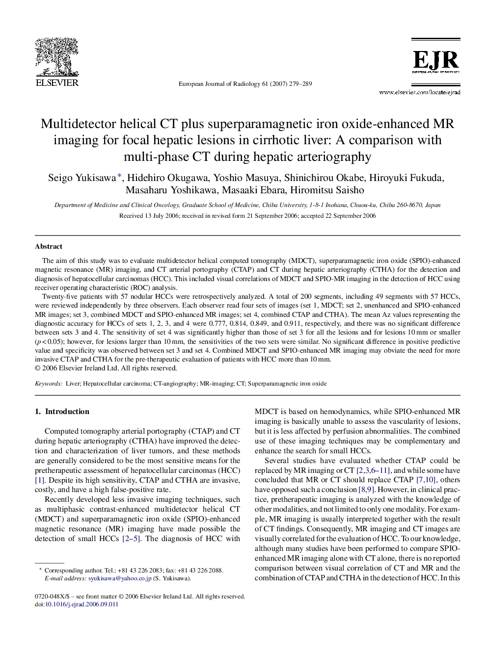
Multidetector helical CT plus superparamagnetic iron oxide-enhanced MR imaging for focal hepatic lesions in cirrhotic liver: A comparison with multi-phase CT during hepatic arteriography
دانلود مقاله + سفارش ترجمه
دانلود مقاله ISI انگلیسی
رایگان برای ایرانیان
کلمات کلیدی
موضوعات مرتبط
علوم پزشکی و سلامت
پزشکی و دندانپزشکی
رادیولوژی و تصویربرداری
پیش نمایش صفحه اول مقاله

چکیده انگلیسی
Twenty-five patients with 57 nodular HCCs were retrospectively analyzed. A total of 200 segments, including 49 segments with 57 HCCs, were reviewed independently by three observers. Each observer read four sets of images (set 1, MDCT; set 2, unenhanced and SPIO-enhanced MR images; set 3, combined MDCT and SPIO-enhanced MR images; set 4, combined CTAP and CTHA). The mean Az values representing the diagnostic accuracy for HCCs of sets 1, 2, 3, and 4 were 0.777, 0.814, 0.849, and 0.911, respectively, and there was no significant difference between sets 3 and 4. The sensitivity of set 4 was significantly higher than those of set 3 for all the lesions and for lesions 10 mm or smaller (p < 0.05); however, for lesions larger than 10 mm, the sensitivities of the two sets were similar. No significant difference in positive predictive value and specificity was observed between set 3 and set 4. Combined MDCT and SPIO-enhanced MR imaging may obviate the need for more invasive CTAP and CTHA for the pre-therapeutic evaluation of patients with HCC more than 10 mm.
ناشر
Database: Elsevier - ScienceDirect (ساینس دایرکت)
Journal: European Journal of Radiology - Volume 61, Issue 2, February 2007, Pages 279-289
Journal: European Journal of Radiology - Volume 61, Issue 2, February 2007, Pages 279-289
نویسندگان
Seigo Yukisawa, Hidehiro Okugawa, Yoshio Masuya, Shinichirou Okabe, Hiroyuki Fukuda, Masaharu Yoshikawa, Masaaki Ebara, Hiromitsu Saisho,