| کد مقاله | کد نشریه | سال انتشار | مقاله انگلیسی | نسخه تمام متن |
|---|---|---|---|---|
| 4236239 | 1282893 | 2007 | 12 صفحه PDF | دانلود رایگان |
عنوان انگلیسی مقاله ISI
Ãchographie des nerfs périphériques - Deuxième partie : membre inférieur
دانلود مقاله + سفارش ترجمه
دانلود مقاله ISI انگلیسی
رایگان برای ایرانیان
کلمات کلیدی
موضوعات مرتبط
علوم پزشکی و سلامت
پزشکی و دندانپزشکی
رادیولوژی و تصویربرداری
پیش نمایش صفحه اول مقاله

چکیده انگلیسی
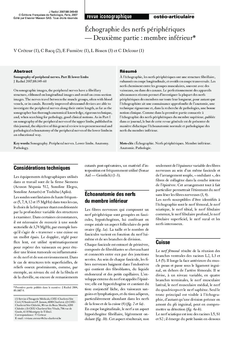
On sonographic images, the peripheral nerves have a fibrillary structure, ribboned on longitudinal images and ovoid on cross-section images. The nerves travel between the muscle groups, often with blood vessels, or in canals. Recently improved ultrasound devices are able to investigate the peripheral nerves along their entire length, as far as the sonographer has thorough anatomical knowledge, rigorous technique, and, when searching for pathology, good clinical notions. As in Part I on sonography of the peripheral nerves of the upper limbs, published in this journal, the objective of this general review is to present normal and pathological echoanatomy of the peripheral nerves of the lower limbs in an educational way.
ناشر
Database: Elsevier - ScienceDirect (ساینس دایرکت)
Journal: Journal de Radiologie - Volume 88, Issue 3, Part 1, March 2007, Pages 349-360
Journal: Journal de Radiologie - Volume 88, Issue 3, Part 1, March 2007, Pages 349-360
نویسندگان
V. Créteur, C. Bacq, E. Fumière, L. Bissen, C. Delcour,