| کد مقاله | کد نشریه | سال انتشار | مقاله انگلیسی | نسخه تمام متن |
|---|---|---|---|---|
| 5696995 | 1410289 | 2017 | 13 صفحه PDF | دانلود رایگان |
عنوان انگلیسی مقاله ISI
Magnetic resonance imaging basics for the prostate brachytherapist
ترجمه فارسی عنوان
اصول اولیه تصویربرداری رزونانس مغناطیسی برای پروتز مغز تراشه
دانلود مقاله + سفارش ترجمه
دانلود مقاله ISI انگلیسی
رایگان برای ایرانیان
کلمات کلیدی
موضوعات مرتبط
علوم پزشکی و سلامت
پزشکی و دندانپزشکی
تومور شناسی
چکیده انگلیسی
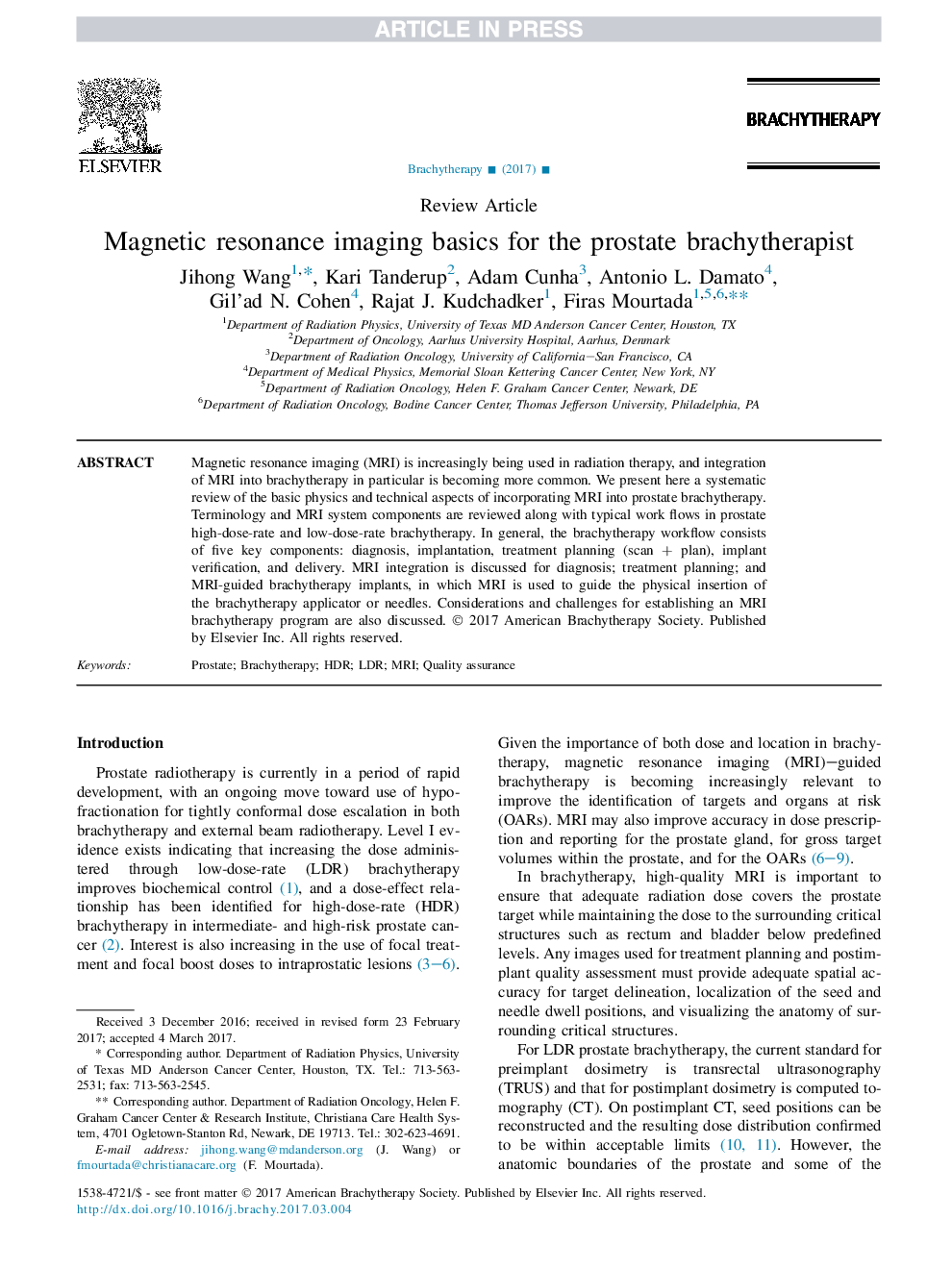
Magnetic resonance imaging (MRI) is increasingly being used in radiation therapy, and integration of MRI into brachytherapy in particular is becoming more common. We present here a systematic review of the basic physics and technical aspects of incorporating MRI into prostate brachytherapy. Terminology and MRI system components are reviewed along with typical work flows in prostate high-dose-rate and low-dose-rate brachytherapy. In general, the brachytherapy workflow consists of five key components: diagnosis, implantation, treatment planning (scan + plan), implant verification, and delivery. MRI integration is discussed for diagnosis; treatment planning; and MRI-guided brachytherapy implants, in which MRI is used to guide the physical insertion of the brachytherapy applicator or needles. Considerations and challenges for establishing an MRI brachytherapy program are also discussed.
ناشر
Database: Elsevier - ScienceDirect (ساینس دایرکت)
Journal: Brachytherapy - Volume 16, Issue 4, JulyâAugust 2017, Pages 715-727
Journal: Brachytherapy - Volume 16, Issue 4, JulyâAugust 2017, Pages 715-727
نویسندگان
Jihong Wang, Kari Tanderup, Adam Cunha, Antonio L. Damato, Gil'ad N. Cohen, Rajat J. Kudchadker, Firas Mourtada,
