| کد مقاله | کد نشریه | سال انتشار | مقاله انگلیسی | نسخه تمام متن |
|---|---|---|---|---|
| 9390198 | 1282749 | 2005 | 5 صفحه PDF | دانلود رایگان |
عنوان انگلیسی مقاله ISI
Unusual Sonographic Feature of Congenital Pulmonary Sequestration
دانلود مقاله + سفارش ترجمه
دانلود مقاله ISI انگلیسی
رایگان برای ایرانیان
کلمات کلیدی
موضوعات مرتبط
علوم پزشکی و سلامت
پزشکی و دندانپزشکی
رادیولوژی و تصویربرداری
پیش نمایش صفحه اول مقاله

چکیده انگلیسی
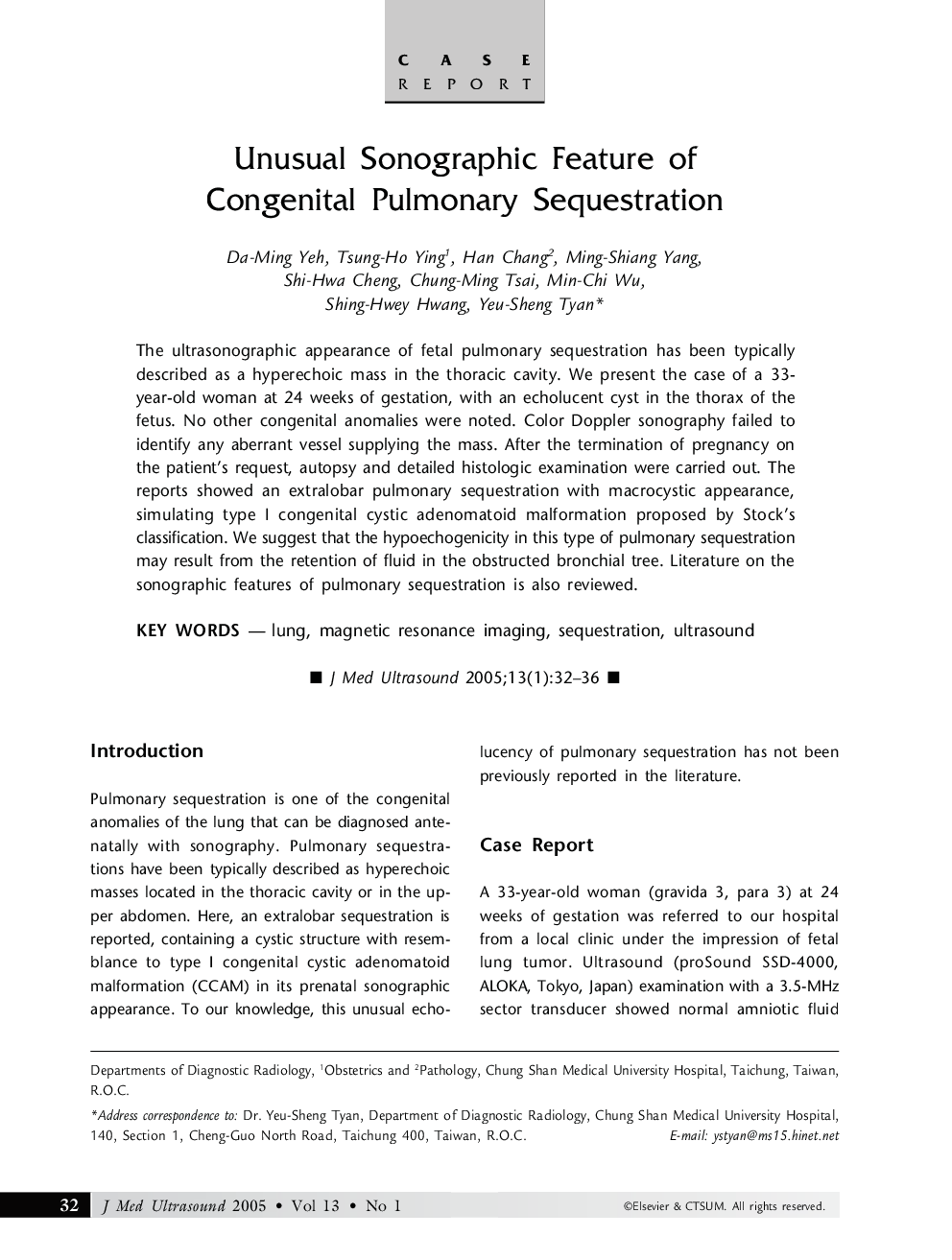
The ultrasonographic appearance of fetal pulmonary sequestration has been typically described as a hyperechoic mass in the thoracic cavity. We present the case of a 33-year-old woman at 24 weeks of gestation, with an echolucent cyst in the thorax of the fetus. No other congenital anomalies were noted. Color Doppler sonography failed to identify any aberrant vessel supplying the mass. After the termination of pregnancy on the patient's request, autopsy and detailed histologic examination were carried out. The reports showed an extralobar pulmonary sequestration with macrocystic appearance, simulating type I congenital cystic adenomatoid malformation proposed by Stock's classification. We suggest that the hypoechogenicity in this type of pulmonary sequestration may result from the retention of fluid in the obstructed bronchial tree. Literature on the sonographic features of pulmonary sequestration is also reviewed.
ناشر
Database: Elsevier - ScienceDirect (ساینس دایرکت)
Journal: Journal of Medical Ultrasound - Volume 13, Issue 1, 2005, Pages 32-36
Journal: Journal of Medical Ultrasound - Volume 13, Issue 1, 2005, Pages 32-36
نویسندگان
Da-Ming Yeh, Ming-Shiang Yang, Shi-Hwa Cheng, Chung-Ming Tsai, Min-Chi Wu, Shing-Hwey Hwang, Yeu-Sheng Tyan, Tsung-Ho Ying, Han Chang,