| کد مقاله | کد نشریه | سال انتشار | مقاله انگلیسی | نسخه تمام متن |
|---|---|---|---|---|
| 10097650 | 1609881 | 2005 | 6 صفحه PDF | دانلود رایگان |
عنوان انگلیسی مقاله ISI
Visualization of femorotibial contact in total knee arthroplasty using X-ray fluoroscopy
دانلود مقاله + سفارش ترجمه
دانلود مقاله ISI انگلیسی
رایگان برای ایرانیان
کلمات کلیدی
موضوعات مرتبط
علوم پزشکی و سلامت
پزشکی و دندانپزشکی
رادیولوژی و تصویربرداری
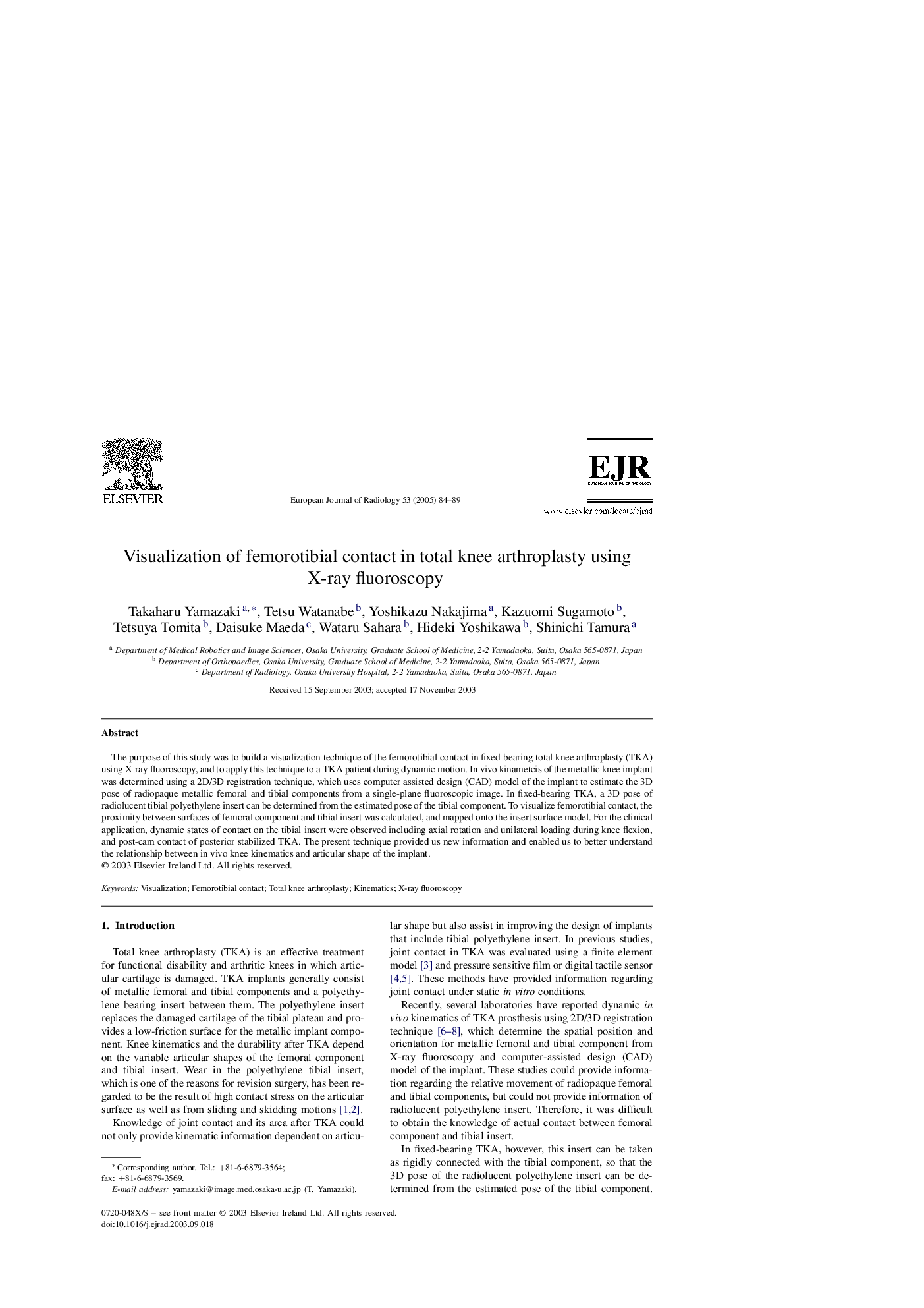
پیش نمایش صفحه اول مقاله

چکیده انگلیسی
The purpose of this study was to build a visualization technique of the femorotibial contact in fixed-bearing total knee arthroplasty (TKA) using X-ray fluoroscopy, and to apply this technique to a TKA patient during dynamic motion. In vivo kinametcis of the metallic knee implant was determined using a 2D/3D registration technique, which uses computer assisted design (CAD) model of the implant to estimate the 3D pose of radiopaque metallic femoral and tibial components from a single-plane fluoroscopic image. In fixed-bearing TKA, a 3D pose of radiolucent tibial polyethylene insert can be determined from the estimated pose of the tibial component. To visualize femorotibial contact, the proximity between surfaces of femoral component and tibial insert was calculated, and mapped onto the insert surface model. For the clinical application, dynamic states of contact on the tibial insert were observed including axial rotation and unilateral loading during knee flexion, and post-cam contact of posterior stabilized TKA. The present technique provided us new information and enabled us to better understand the relationship between in vivo knee kinematics and articular shape of the implant.
ناشر
Database: Elsevier - ScienceDirect (ساینس دایرکت)
Journal: European Journal of Radiology - Volume 53, Issue 1, January 2005, Pages 84-89
Journal: European Journal of Radiology - Volume 53, Issue 1, January 2005, Pages 84-89
نویسندگان
Takaharu Yamazaki, Tetsu Watanabe, Yoshikazu Nakajima, Kazuomi Sugamoto, Tetsuya Tomita, Daisuke Maeda, Wataru Sahara, Hideki Yoshikawa, Shinichi Tamura,