| کد مقاله | کد نشریه | سال انتشار | مقاله انگلیسی | نسخه تمام متن |
|---|---|---|---|---|
| 9230352 | 1203629 | 2005 | 11 صفحه PDF | دانلود رایگان |
عنوان انگلیسی مقاله ISI
Melanoma Cells Express Elevated Levels of Phosphorylated Histone H2AX Foci
دانلود مقاله + سفارش ترجمه
دانلود مقاله ISI انگلیسی
رایگان برای ایرانیان
کلمات کلیدی
موضوعات مرتبط
علوم پزشکی و سلامت
پزشکی و دندانپزشکی
امراض پوستی
پیش نمایش صفحه اول مقاله

چکیده انگلیسی
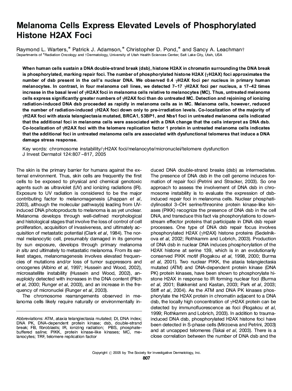
When human cells sustain a DNA double-strand break (dsb), histone H2AX in chromatin surrounding the DNA break is phosphorylated, marking repair foci. The number of phosphorylated histone H2AX (γH2AX) foci approximates the number of dsb present in the cell's nuclear DNA. We observed 0.4 γH2AX foci per nucleus in primary human melanocytes. In contrast, in four melanoma cell lines, we detected 7-17 γH2AX foci per nucleus, a 17-42 times increase in the basal level of γH2AX foci in melanoma cells relative to melanocytes (MC). Thus, untreated melanoma cells express significantly greater numbers of γH2AX foci than do untreated MC. Detection and rejoining of ionizing radiation-induced DNA dsb proceeded as rapidly in melanoma cells as in MC. Melanoma cells, however, reduced the number of radiation-induced γH2AX foci down only to pre-irradiation levels. Co-localization of the majority of γH2AX foci with ataxia telangiectasia mutated, BRCA1, 53BP1, and Nbs1 foci in untreated melanoma cells indicated that the additional foci in melanoma cells were associated with a DNA change that the cells interpret as DNA dsb. Co-localization of γH2AX foci with the telomere replication factor 1 protein in untreated melanoma cells indicates that the additional foci in untreated melanoma cells are associated with dysfunctional telomeres that induce a DNA damage stress response.
ناشر
Database: Elsevier - ScienceDirect (ساینس دایرکت)
Journal: Journal of Investigative Dermatology - Volume 124, Issue 4, April 2005, Pages 807-817
Journal: Journal of Investigative Dermatology - Volume 124, Issue 4, April 2005, Pages 807-817
نویسندگان
Raymond L. Warters, Patrick J. Adamson, Christopher D. Pond, Sancy A. Leachman,