| کد مقاله | کد نشریه | سال انتشار | مقاله انگلیسی | نسخه تمام متن |
|---|---|---|---|---|
| 9232478 | 1204428 | 2005 | 6 صفحه PDF | دانلود رایگان |
عنوان انگلیسی مقاله ISI
Imaging Melanoma in a Murine Model Using Reflectance-Mode Confocal Scanning Laser Microscopy and Polarized Light Imaging
دانلود مقاله + سفارش ترجمه
دانلود مقاله ISI انگلیسی
رایگان برای ایرانیان
کلمات کلیدی
موضوعات مرتبط
علوم پزشکی و سلامت
پزشکی و دندانپزشکی
امراض پوستی
پیش نمایش صفحه اول مقاله

چکیده انگلیسی
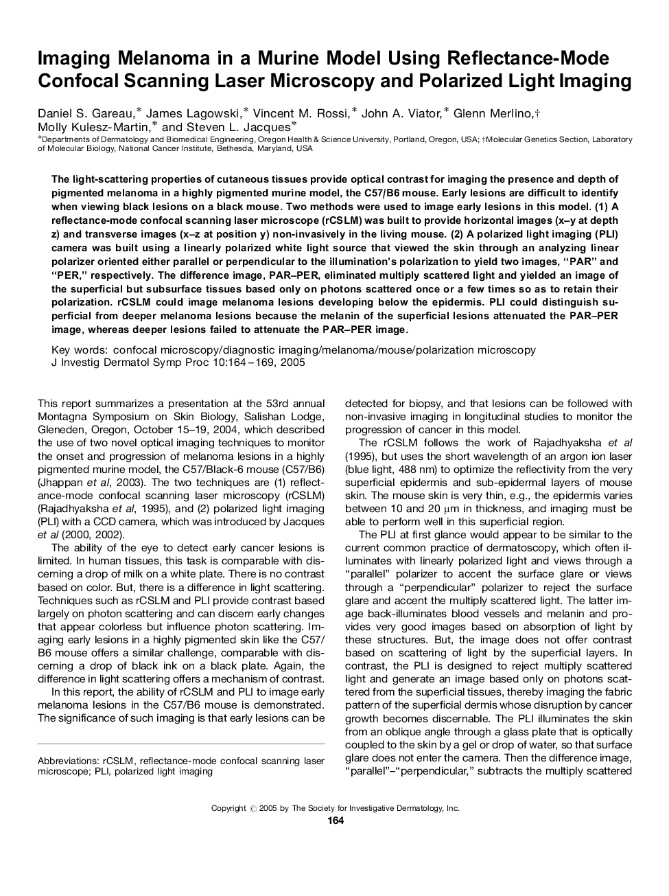
The light-scattering properties of cutaneous tissues provide optical contrast for imaging the presence and depth of pigmented melanoma in a highly pigmented murine model, the C57/B6 mouse. Early lesions are difficult to identify when viewing black lesions on a black mouse. Two methods were used to image early lesions in this model. (1) A reflectance-mode confocal scanning laser microscope (rCSLM) was built to provide horizontal images (x-y at depth z) and transverse images (x-z at position y) non-invasively in the living mouse. (2) A polarized light imaging (PLI) camera was built using a linearly polarized white light source that viewed the skin through an analyzing linear polarizer oriented either parallel or perpendicular to the illumination's polarization to yield two images, “PAR” and “PER,” respectively. The difference image, PAR-PER, eliminated multiply scattered light and yielded an image of the superficial but subsurface tissues based only on photons scattered once or a few times so as to retain their polarization. rCSLM could image melanoma lesions developing below the epidermis. PLI could distinguish superficial from deeper melanoma lesions because the melanin of the superficial lesions attenuated the PAR-PER image, whereas deeper lesions failed to attenuate the PAR-PER image.
ناشر
Database: Elsevier - ScienceDirect (ساینس دایرکت)
Journal: Journal of Investigative Dermatology Symposium Proceedings - Volume 10, Issue 2, November 2005, Pages 164-169
Journal: Journal of Investigative Dermatology Symposium Proceedings - Volume 10, Issue 2, November 2005, Pages 164-169
نویسندگان
Daniel S. Gareau, James Lagowski, Vincent M. Rossi, John A. Viator, Glenn Merlino, Molly Kulesz-Martin, Steven L. Jacques,